 Indietro
Microbial Cell Structure and Function: Study Notes
Guida di studio - Note intelligenti
 Appunti personalizzati basati sui tuoi materiali, ampliati con definizioni chiave, esempi e contesto.
Microbial Cell Structure and Function
Overview of Microbial Cell Structures
Microbial cells possess a variety of structures that contribute to their survival, adaptation, and interaction with their environment. These structures include the cell envelope, cell wall, capsules, fimbriae, pili, flagella, inclusions, and endospores. Each component plays a specific role in cellular physiology and pathogenicity.
Cell Envelope/Membrane: Composed of phospholipid bilayers, it regulates interactions with the environment.
Cell Wall: Provides structural support; peptidoglycan is thick in Gram-positive and thin in Gram-negative bacteria.
Capsules: Polysaccharide layers that enhance virulence by preventing phagocytosis.
Fimbriae: Facilitate attachment to surfaces.
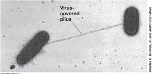
Pili: Involved in conjugation and genetic exchange.
Flagella: Enable motility.
Inclusions: Serve as storage and energy reserves.
Endospores: Allow survival under harsh conditions.
The Cell Envelope
Structure and Components
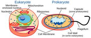
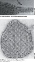
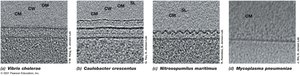
The cell envelope is a series of layered structures that surround the cytoplasm and mediate interactions with the environment. All cells have a cytoplasmic membrane, most bacteria have a cell wall, and Gram-negative bacteria possess an additional outer membrane. Some bacteria also have S-layers composed of protein or glycoprotein.
Gram-Positive vs. Gram-Negative Bacteria
Bacteria are classified based on their cell wall structure, which is revealed by the Gram stain:
Gram-Positive: Thick peptidoglycan layer, no outer membrane.
Gram-Negative: Thin peptidoglycan layer, outer membrane present.
The Cytoplasmic (Cell) Membrane
Structure and Function
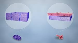
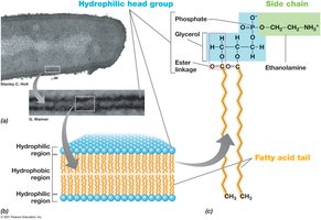
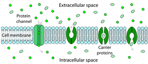
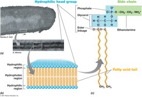
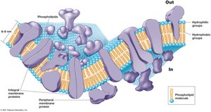
The cytoplasmic membrane is a phospholipid bilayer containing embedded proteins. It is 8–10 nm wide and separates the cytoplasm from the environment. Its main function is selective permeability, allowing nutrients in and waste out. Membrane proteins facilitate transport and energy metabolism.
Phospholipid Bilayer Details
Hydrophobic Tails: Fatty acids oriented inward.
Hydrophilic Heads: Glycerol, phosphate, and functional groups exposed to the environment or cytoplasm.
Membrane Proteins
Integral (Transmembrane) Proteins: Span the membrane.
Peripheral Proteins: Loosely attached to the membrane surface.
Variations Across Domains
Archaeal membranes differ from bacterial and eukaryotic membranes:
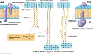
Archaea: Ether linkages in phospholipids, isoprene side chains, can form monolayers.
Bacteria/Eukarya: Ester linkages, fatty acid side chains, always bilayers.
Other Functions of the Cytoplasmic Membrane
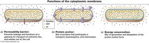
Permeability Barrier: Prevents leakage and acts as a gateway for nutrient and waste transport.
Protein Anchor: Site for proteins involved in transport, bioenergetics, and chemotaxis.
Energy Conservation: Site of generation and dissipation of the proton motive force.
The Cell Wall
Function and Composition
The cell wall provides rigidity, maintains cell shape, and protects against osmotic lysis. Most bacteria are classified as Gram-positive or Gram-negative based on cell wall structure. Acid-fast bacteria have unique, waxy cell walls.
Peptidoglycan Structure
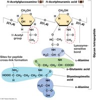
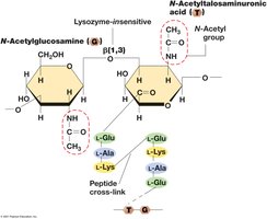
Peptidoglycan is a rigid polysaccharide layer unique to bacteria. It consists of a sugar backbone (alternating N-acetylglucosamine and N-acetylmuramic acid) joined by β-1,4 linkages, with short peptides attached to N-acetylmuramic acid. Amino acid composition varies by species.
Gram-Positive Cell Wall
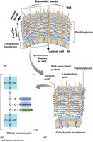
Thick peptidoglycan layer (up to 90% of wall, 15+ layers).
Stabilized by peptide cross-links and interbridges (e.g., glycine bridges in Staphylococcus aureus).
Teichoic acids embedded in the wall; lipoteichoic acids link to membrane lipids.
Susceptible to lysozyme and penicillin.
Gram-Negative Cell Wall
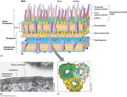
Thin peptidoglycan layer (single layer), located in the periplasmic space.
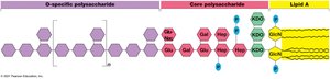
Outer membrane contains lipopolysaccharide (LPS), porins, and Braun lipoprotein.
Lipid A component of LPS acts as an endotoxin.
Archaeal Cell Walls
Lack peptidoglycan and outer membrane.
Most have S-layers (protein shells); some methanogens have pseudomurein (alternating N-acetylglucosamine and N-acetyltalosaminuronic acid, β-1,3 linkages).
Resistant to lysozyme and penicillin.
Diversity of Cell Envelope Structures
S-Layers: Paracrystalline protein or glycoprotein layers, always outermost if present, provide strength and protection.
Wall-less Bacteria/Archaea: Some (e.g., Mycoplasma, Thermoplasma) lack cell walls but have tough membranes with sterols.
Cell Surface Structures
Capsules and Slime Layers
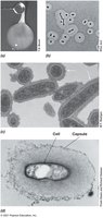
Capsules are tightly attached polysaccharide coats, while slime layers are loosely attached and easily deformed. Both assist in surface attachment, biofilm formation, infectivity, and protection from desiccation.
Pili and Fimbriae
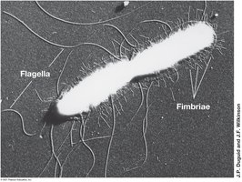
Pili: Thin protein filaments for attachment, conjugation, and motility (e.g., twitching).
Fimbriae: Shorter, more numerous, mediate attachment to surfaces.
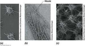
Hamus/Hami (Archaea)
Unique grappling hook-like structures in some Archaea, aiding in attachment and biofilm formation.
Cell Inclusions
Types and Functions
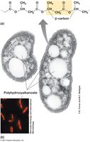
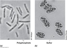
Inclusions serve as energy reserves or reservoirs for carbon, phosphorus, sulfur, or other minerals. They are enclosed by thin protein membranes, reducing osmotic stress.
Carbon Storage: Poly-β-hydroxybutyric acid (PHB), poly-β-hydroxyalkanoate (PHA), and glycogen.
Polyphosphate Granules: Store inorganic phosphate.
Sulfur Granules: Store elemental sulfur, oxidized to sulfate.
Carbonate Minerals: Biomineralization of barium, strontium, magnesium.
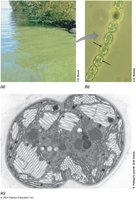
Gas Vesicles: Confer buoyancy, especially in cyanobacteria.
Magnetosomes: Allow orientation within magnetic fields (magnetotaxis).
Endospores
Formation and Function
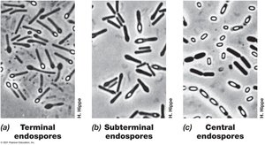
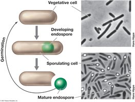
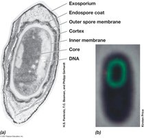
Endospores are highly differentiated, dormant cells formed by some Gram-positive bacteria (e.g., Bacillus, Clostridium). They are resistant to heat, radiation, chemicals, and desiccation, allowing survival in harsh conditions. Endospores are ideal for dispersal and can remain dormant for years, germinating rapidly when conditions improve.
Endospore Structure
Contains dipicolinic acid and is enriched in Ca2+.
Core contains small acid-soluble spore proteins (SASPs) that protect DNA and serve as energy sources during outgrowth.
Flagella, Archaella, and Motility
Flagella and Swimming Motility
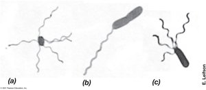
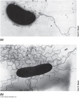
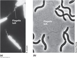
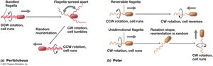
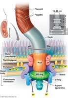
Flagella (in Bacteria) and archaella (in Archaea) are rotating appendages that enable swimming. Arrangements include polar, lophotrichous, amphitrichous, and peritrichous. Movement is powered by the proton motive force.
Surface Motility
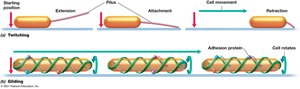
Twitching Motility: Pili extend, attach, and retract to pull the cell forward (requires ATP hydrolysis).
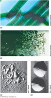
Gliding Motility: Smooth movement along surfaces without external appendages, involving protein tracks and motors.
Chemotaxis and Other Forms of Taxis
Chemotaxis
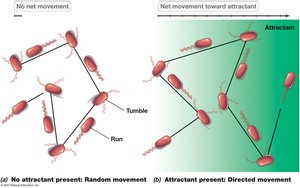
Chemotaxis is the directed movement of cells in response to chemical gradients. Bacteria like E. coli exhibit "run and tumble" behavior, adjusting movement based on temporal changes in attractant or repellent concentration. Chemoreceptors sense these changes, leading to a biased random walk toward favorable environments.
Other Forms of Taxis
Osmotaxis: Response to ionic strength.
Hydrotaxis: Response to water.
Aerotaxis: Response to oxygen concentration.
Phototaxis: Response to light, optimizing position for phototrophic growth.
Magnetotaxis: Orientation and movement along magnetic field lines, often linked to aerotaxis.
Endosymbiotic Origin of Organelles
Endosymbiotic Hypothesis
The endosymbiotic hypothesis proposes that mitochondria and chloroplasts originated from free-living bacteria that established symbiotic relationships with ancestral eukaryotic cells. Evidence includes the presence of circular DNA and bacterial-like ribosomes in these organelles.
Mitochondria descended from respiratory bacteria.
Chloroplasts descended from phototrophic bacteria.
Summary Table: Key Differences in Cell Envelope Structures
Feature | Gram-Positive Bacteria | Gram-Negative Bacteria | Archaea |
|---|---|---|---|
Peptidoglycan | Thick, multi-layered | Thin, single-layered | Absent (may have pseudomurein or S-layer) |
Outer Membrane | Absent | Present (contains LPS) | Absent |
Teichoic Acids | Present | Absent | Absent |
Lipid Composition | Ester-linked fatty acids | Ester-linked fatty acids | Ether-linked isoprenoids |
Example | Bacillus, Staphylococcus | Escherichia coli | Halobacterium, Methanogens |
