 Indietro
Microbial Diseases of the Skin and Wounds – Study Notes
Guida di studio - Note intelligenti
 Appunti personalizzati basati sui tuoi materiali, ampliati con definizioni chiave, esempi e contesto.
Microbial Diseases of the Skin and Wounds
Anatomy and Defense Mechanisms of the Skin
The skin is the largest organ of the human body and serves as a primary barrier against microbial invasion. It consists of two main layers: the epidermis (outer layer) and the dermis (inner layer). The outermost layer is composed of dead, keratinized cells, making it impenetrable to most microbes. The skin also prevents excessive water loss, regulates temperature, and is involved in sensory phenomena. Its surface is salty and contains sebum, favoring the growth of halophilic and sebum-resistant normal flora.
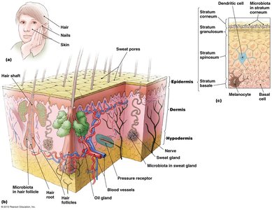
Normal Flora: Includes bacteria (e.g., Staphylococcus, Micrococcus, diphtheroids) and fungi (e.g., Malassezia).
These microbes are generally harmless but can cause disease if the skin barrier is breached or the immune system is suppressed.
Wounds and Microbial Invasion
Wounds are traumas to any tissue of the body (cuts, scrapes, burns, bites, surgery) that allow microbes to infect deeper tissues. While body defenses usually eliminate infections, some wounds can result in severe or fatal diseases.
Bacterial Diseases of the Skin
Folliculitis, Furuncles, and Carbuncles
Folliculitis is an infection of the hair follicle, often presenting as a pimple. When it occurs at the eyelid base, it is called a sty. Spread of infection can lead to furuncles (boils), and when multiple furuncles coalesce, they form carbuncles (usually in thick skin areas).
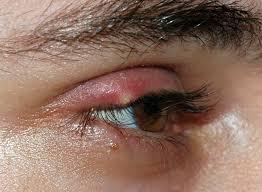


Pathogen: Most commonly caused by Staphylococcus species (Gram-positive cocci in clusters), especially S. aureus and S. epidermidis.
Virulence Factors: S. aureus produces beta-lactamase and toxins, making it more pathogenic than S. epidermidis.
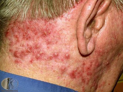
Diagnosis: Isolation of Gram-positive cocci in clusters from pus.
Treatment: Dicloxacillin (drug of choice); vancomycin for resistant strains.
Prevention: Hand antisepsis and proper hospital procedures to minimize MRSA infections.
Staphylococcal Scalded Skin Syndrome (SSSS)
SSSS is caused by certain strains of S. aureus that produce exfoliative toxins, leading to widespread sloughing of the skin. The dermis remains unaffected, so no scarring occurs. The disease primarily affects infants and is transmitted person-to-person.
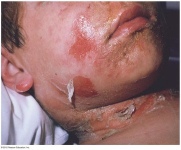
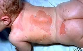
Diagnosis: Characteristic sloughing of skin.
Treatment: Administration of antimicrobial drugs.
Prevention: Difficult due to widespread presence of S. aureus.
Impetigo (Pyoderma) and Erysipelas
Both are superficial skin infections. Impetigo is characterized by vesicles and is most common in children, while erysipelas affects the elderly and involves deeper dermal layers.

Pathogens: Mainly S. aureus, but also Streptococcus pyogenes (Group A Strep).
Virulence Factors: M protein, hyaluronic acid, pyrogenic toxins.
Diagnosis: Presence of vesicles (impetigo).
Treatment: Topical mupirocin and oral clindamycin or doxycycline for impetigo; penicillin for erysipelas.
Prevention: Proper hygiene and cleanliness.
Necrotizing Fasciitis (Flesh-Eating Disease)
This rapidly progressing infection is most often caused by Streptococcus pyogenes (Group A Strep). The bacteria produce enzymes and toxins that destroy tissues, leading to high mortality if not treated promptly.
Diagnosis: Early diagnosis is difficult due to nonspecific symptoms.
Treatment: Clindamycin and penicillin or broad-spectrum antibiotics.
Acne
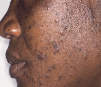
Acne is commonly caused by Propionibacterium acnes, a Gram-positive, rod-shaped bacterium that is part of the normal skin microbiota. Acne typically begins in adolescence due to increased sebum production.
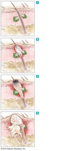

Treatment: Antimicrobial drugs, exfoliating agents, Accutane for severe cases, and blue-light therapy.
Pseudomonas Infection
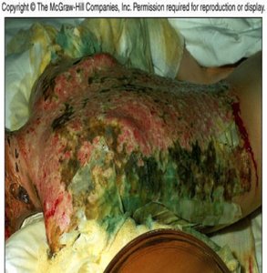
Pseudomonas aeruginosa is a Gram-negative bacterium found in soil and moist environments. It is an opportunistic pathogen, especially in burn victims, and produces a greenish pigment (pyocyanin) in massive infections.
Virulence Factors: Adhesins, capsule, toxins, enzymes, fimbriae, pyocyanin.
Treatment: Difficult due to multidrug resistance; sulfadiazine and piperacillin are used.
Cutaneous Anthrax
Caused by Bacillus anthracis, cutaneous anthrax occurs when spores enter the skin, forming a black, painless ulcer called an eschar. It is treated with doxycycline or ciprofloxacin.
Gas Gangrene
Gas gangrene is caused by Clostridium species (especially C. perfringens). It is characterized by ischemia, necrosis, blackening of skin, and gas bubbles. Rapid treatment is crucial due to high mortality.
Treatment: Surgical removal of dead tissue, antitoxin, and penicillin.
Viral Diseases of the Skin
Poxviruses
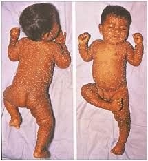
Poxviruses cause diseases such as smallpox, orf, cowpox, and monkeypox. Smallpox was eradicated globally through vaccination. The disease progresses through macule, papule, vesicle, pustule, crust, and scar stages.
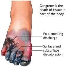
Prevention: Vaccination (discontinued after eradication of smallpox).
Herpes Infections
Herpes simplex viruses (HSV-1 and HSV-2) cause slow-spreading, painful skin lesions. The virus can become latent in nerve ganglia and reactivate later.
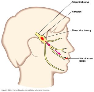
Treatment: Antiviral drugs (acyclovir, penciclovir) control but do not cure the infection.
Chickenpox and Shingles
Both are caused by the varicella-zoster virus (VZV). Chickenpox presents as a widespread rash, while shingles is a localized, painful rash along a nerve in adults due to reactivation of latent VZV.
Prevention: Vaccine available for both diseases.
Warts
Warts are benign epithelial growths caused by various papillomaviruses. They are usually harmless but can be oncogenic. Transmission occurs via direct contact and fomites.
Rubella (German Measles)
Rubella is a mild rash disease in children but can cause severe congenital defects if contracted during pregnancy. It is caused by the rubella virus and prevented by vaccination.
Measles (Rubeola)
Measles is a highly contagious viral disease characterized by Koplik’s spots. It is prevented by the MMR vaccine.
Fungal Diseases (Mycoses) of the Skin
Superficial Mycoses
These infections occur on the hair, nails, and outer skin layers. Examples include ringworm (dermatophytes), piedra (nodules on hair shaft), and pityriasis versicolor (scaly, pigmented patches).
Diagnosis: Appearance, green color under UV light (pityriasis).
Treatment: Topical or oral antifungal drugs.
Cutaneous Mycoses
Dermatophytoses are cutaneous infections caused by dermatophytes. They can be diagnosed by clinical observation and confirmed with KOH preparation of skin or nail samples.
Treatment: Topical agents for limited infections; oral drugs for widespread infections.
