 Indietro
Microbial Growth and Its Control: Structured Study Notes
Guida di studio - Note intelligenti
 Appunti personalizzati basati sui tuoi materiali, ampliati con definizioni chiave, esempi e contesto.
Microbial Growth and Its Control
4.1 Feeding the Microbe: Cell Nutrition
Microbial growth requires a balanced supply of nutrients, which are classified as macronutrients and micronutrients. Understanding the chemical composition of microbial cells is essential for designing appropriate growth media and studying microbial physiology.
Macronutrients: Elements required in large amounts, including carbon (C), oxygen (O), nitrogen (N), hydrogen (H), phosphorus (P), and sulfur (S), which together make up about 96% of the dry weight of a bacterial cell.
Micronutrients: Elements required in trace amounts, such as iron (Fe), manganese (Mn), zinc (Zn), and copper (Cu), often serving as enzyme cofactors.
Growth Factors: Organic compounds like vitamins, amino acids, purines, and pyrimidines that some microbes cannot synthesize and must obtain from the environment.
Heterotrophs obtain carbon from organic compounds, while autotrophs use carbon dioxide as their carbon source.
Nitrogen sources include ammonia (NH3), nitrate (NO3-), nitrogen gas (N2), and organic nitrogen compounds.
Phosphorus is usually assimilated as inorganic phosphate (PO43-), and sulfur as sulfate (SO42-), sulfide (H2S), or organics.
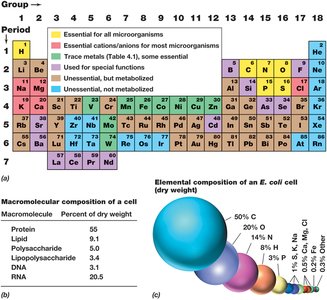
Additional info: The periodic table in the image highlights essential elements for microbial life, and the pie chart shows the relative abundance of macromolecules in a typical bacterial cell.
4.2 Growth Media and Laboratory Culture
Microorganisms are cultivated in the laboratory using nutrient solutions called culture media, which can be tailored to meet the specific nutritional requirements of different microbes.
Defined media: Contain precise amounts of pure chemicals; the exact composition is known.
Complex media: Contain extracts or digests of natural products (e.g., yeast extract, peptone); composition is not precisely known.
Selective media: Contain compounds that inhibit the growth of some microbes while allowing others to grow.
Differential media: Contain indicators that reveal differences between microbial species based on metabolic reactions.
Solid media are prepared by adding agar, allowing the formation of isolated colonies, which can be used to assess colony morphology and purity.
Aseptic technique is essential to prevent contamination during microbial transfers.
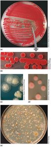
Additional info: The image shows various colony morphologies, which can aid in the identification of microorganisms.
4.3 Microscopic Counts of Microbial Cell Numbers
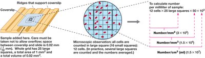
Microscopic cell counting is a direct method for estimating the number of microbial cells in a sample, using specialized counting chambers.
Counting chambers (e.g., Petroff–Hausser chamber) allow enumeration of cells in a defined volume.
Limitations: Cannot distinguish live from dead cells without special stains; small cells and debris may be miscounted; time-consuming and less precise for low concentrations.
Staining techniques (e.g., DAPI, fluorescent stains) can differentiate live/dead cells and provide phylogenetic information.
4.4 Viable Counting of Microbial Cell Numbers
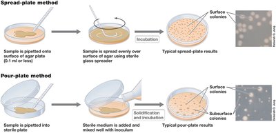
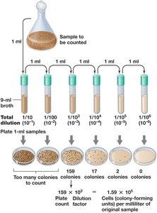
Viable (plate) counts estimate the number of living, reproducing cells in a sample by counting colonies formed on solid media.
Spread-plate method: Sample is spread over the surface of an agar plate.
Pour-plate method: Sample is mixed with molten agar and poured into a plate.
Results are reported as colony-forming units (CFU) to account for cell clumps.
Serial dilutions are used to obtain countable plates (30–300 colonies).
Applications: Used in food, water, and clinical microbiology for quantifying specific microbes.
Great plate count anomaly: Direct counts often reveal more cells than can be cultured, due to differing growth requirements.
4.5 Turbidimetric Measures of Microbial Cell Numbers
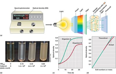
Turbidity measurements estimate cell numbers based on the cloudiness of a cell suspension, using a spectrophotometer to measure optical density (OD).
Optical density (OD): Proportional to cell concentration within certain limits.
Standard curve: Must be established to relate OD to actual cell numbers.
Advantages: Rapid, non-destructive, and allows repeated measurements.
Limitations: Not suitable for clumped or biofilm-forming microbes.
4.6 Binary Fission and the Microbial Growth Cycle
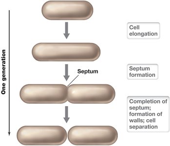
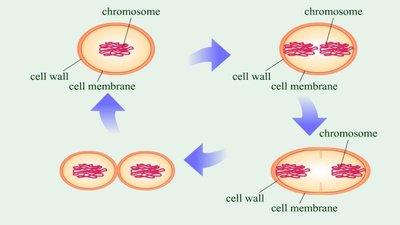
Most bacteria reproduce by binary fission, a process in which a cell grows, duplicates its DNA, and divides into two genetically identical daughter cells. The growth of a microbial population in a closed system follows a characteristic growth curve.
Binary fission: Cell elongates, forms a septum, and divides into two cells.
Generation time: Time required for a population to double in number (e.g., E. coli ~20 min under optimal conditions).
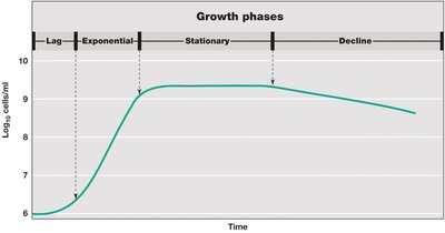
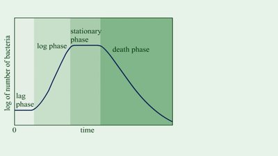
Batch culture: Closed system with fixed volume; growth curve has four phases:
Lag phase: Adaptation, no immediate increase in cell number.
Exponential (log) phase: Cells divide at a constant, maximum rate.
Stationary phase: Growth rate slows/stops due to nutrient depletion or waste accumulation.
Death phase: Cells die at an exponential rate.
4.7 Quantitative Aspects of Microbial Growth
The increase in cell number during exponential growth can be described mathematically:
Exponential growth equation:
Where is the cell number at time t, is the initial cell number, and n is the number of generations.
4.8 Continuous Culture
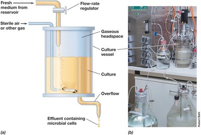
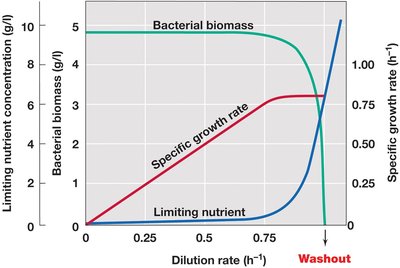
Continuous culture systems, such as the chemostat, maintain microbial populations in exponential growth by continuously adding fresh medium and removing spent medium and cells.
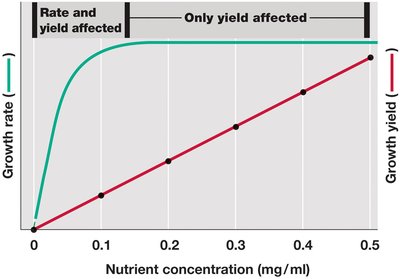
Chemostat: Controls both the specific growth rate (by dilution rate) and cell yield (by limiting nutrient concentration).
Steady state: Cell density and substrate concentration remain constant over time.
Applications: Used to study microbial physiology, ecology, and evolution under controlled conditions.
4.9 Biofilm Growth
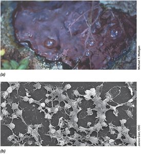
Microbes can grow as free-floating (planktonic) cells or as surface-attached communities called biofilms, which have distinct properties and are important in medical and industrial contexts.
Biofilm: Cells embedded in a self-produced polysaccharide matrix attached to a surface.
Stages of biofilm development: Attachment, colonization, development, and dispersal.
Biofilms are implicated in persistent infections, device contamination, and industrial fouling.

4.10 Alternatives to Binary Fission
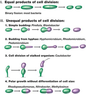
Some bacteria reproduce by mechanisms other than binary fission, such as budding and hyphal growth.
Budding: Unequal cell division producing a daughter cell from a localized site.
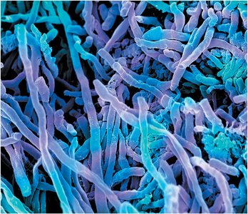
Hyphal growth: Filamentous bacteria (e.g., Streptomyces) grow by extending hyphae, forming mycelia and arthrospores.
4.11 Temperature Classes of Microorganisms
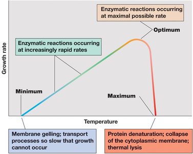
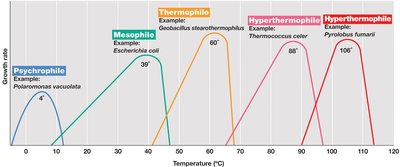
Microorganisms are classified based on their temperature growth optima, which reflect adaptations to their environments.
Cardinal temperatures: Minimum, optimum, and maximum temperatures for growth.
Temperature classes:
Psychrophiles: Optimum ≤ 15°C, found in cold environments.
Mesophiles: Optimum 20–45°C, includes most human-associated microbes.
Thermophiles: Optimum 45–80°C, found in hot environments.
Hyperthermophiles: Optimum >80°C, found in extremely hot habitats.
4.12 Microbial Life in the Cold
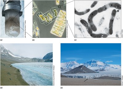
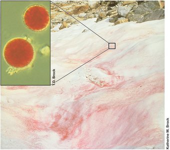
Psychrophilic and psychrotolerant microorganisms have evolved molecular adaptations to thrive in cold environments, such as polar regions and deep oceans.
Psychrophiles: True cold-adapted organisms with optimal growth at ≤15°C.
Psychrotolerant: Can grow at 0°C but have higher optima (20–40°C).
Adaptations: Enzymes with more α-helices, higher unsaturated fatty acids in membranes, cold shock proteins, and cryoprotectants.
4.13 Microbial Life at High Temperatures
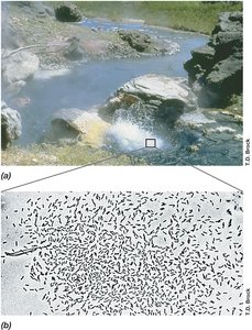
Thermophiles and hyperthermophiles are adapted to hot environments, with specialized proteins and membrane structures for stability at high temperatures.
Thermophiles: Optimum 45–80°C; Hyperthermophiles: Optimum >80°C.
Protein adaptations: Increased ionic bonds, hydrophobic interiors, and stabilizing solutes.
Membrane adaptations: Saturated fatty acids in Bacteria; ether-linked lipids and monolayers in Archaea.
Applications: Thermostable enzymes (e.g., Taq polymerase) are valuable in biotechnology.
4.14 Effects of pH on Microbial Growth
Microorganisms are classified by their pH growth optima, and most maintain a near-neutral cytoplasmic pH regardless of external conditions.
Neutrophiles: Optimum pH 5.5–7.9.
Acidophiles: Optimum pH <5.5; require acidic environments.
Alkaliphiles: Optimum pH ≥8; found in alkaline habitats.
Buffering: Culture media often contain buffers to maintain stable pH.
4.15 Osmolarity and Microbial Growth
Water availability, expressed as water activity (aw), is critical for microbial growth. Microbes are classified by their tolerance to solute concentrations.
Halophiles: Require NaCl for growth; found in marine environments.
Halotolerant: Tolerate some salt but grow best without it.
Extreme halophiles: Require very high salt concentrations.
Osmophiles: Thrive in high-sugar environments; xerophiles: Grow in very dry conditions.
Compatible solutes: Accumulated to maintain positive water balance without disrupting cell function.
4.16 Oxygen and Microbial Growth
Microorganisms vary in their oxygen requirements and tolerance, which is reflected in their metabolism and preferred habitats.
Obligate aerobes: Require oxygen for growth.
Facultative anaerobes: Can grow with or without oxygen.
Microaerophiles: Require reduced oxygen levels.
Aerotolerant anaerobes: Tolerate oxygen but do not use it.
Obligate anaerobes: Oxygen is toxic; grow only in its absence.
Oxygen toxicity: Due to reactive oxygen species (ROS) such as superoxide anion, hydrogen peroxide, and hydroxyl radical.
Protective enzymes: Catalase, peroxidase, superoxide dismutase, and superoxide reductase detoxify ROS.
4.17 General Principles and Microbial Growth Control by Heat
Physical and chemical methods are used to control microbial growth in laboratory and clinical settings.
Decontamination: Making objects safe to handle.
Disinfection: Killing or inhibiting pathogens, not necessarily all microbes.
Heat sterilization: Most common method; effectiveness depends on temperature and exposure time.
Autoclave: Uses steam under pressure (121°C) to sterilize; kills endospores.
Pasteurization: Reduces microbial load in heat-sensitive liquids; does not sterilize.
4.18 Other Physical Control Methods: Radiation and Filtration
Radiation and filtration are alternative methods for controlling microbial growth, especially for heat-sensitive materials.
Ultraviolet (UV) radiation: Damages DNA; used for surface decontamination.
Ionizing radiation: Produces ions and radicals; used for sterilizing medical supplies and food.
Filtration: Removes microbes from liquids and gases using depth or membrane filters.
4.19 Chemical Control of Microbial Growth
Chemical agents are used to kill or inhibit microbes on surfaces, in liquids, and on living tissues.
-cidal agents: Kill microorganisms (e.g., bactericidal, fungicidal).
-static agents: Inhibit growth without killing (e.g., bacteriostatic).
Assaying antimicrobial activity: Minimum inhibitory concentration (MIC) and disk diffusion assays determine effectiveness.
Types of chemical agents:
Sterilants: Destroy all microbes, including endospores.
Disinfectants: Kill most microbes, not necessarily endospores.
Sanitizers: Reduce microbial numbers to safe levels.
Antiseptics: Safe for use on living tissues.
