 Indietro
Microbiology Lab Final Exam Review: Biochemical Tests and Microbial Physiology
Guida di studio - Note intelligenti
 Appunti personalizzati basati sui tuoi materiali, ampliati con definizioni chiave, esempi e contesto.
Phenol Red Sugar Broth
Principle and Interpretation
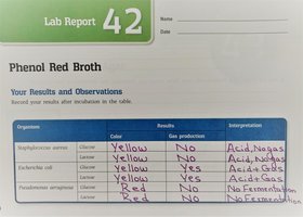
The Phenol Red Sugar Broth test is used to determine the ability of microorganisms to ferment specific carbohydrates, producing acid and/or gas as end products. The medium contains a single carbohydrate (such as glucose or lactose), a pH indicator (phenol red), and a Durham tube to capture gas.
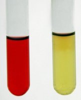
Phenol Red Indicator: Turns yellow in acidic conditions (fermentation), remains red in alkaline or neutral conditions.
Durham Tube: Collects gas produced during fermentation.
Results Interpretation:
Yellow color: Acid production (fermentation)
Yellow with gas bubble: Acid and gas production
Red color: No fermentation
Symbols: A/G (acid and gas), A/- (acid only), K (alkaline, no fermentation)
Substrates: Commonly glucose or lactose.

Triple Sugar Iron (TSI) Agar
Principle and Interpretation
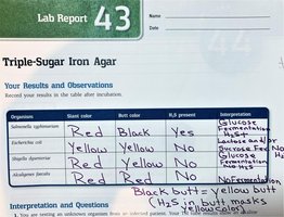
TSI agar is a differential medium used to distinguish bacteria based on their ability to ferment glucose, lactose, and/or sucrose, and to produce hydrogen sulfide (H2S). The medium contains phenol red as a pH indicator and iron salts for H2S detection.
Fermentation Results:
Yellow butt/yellow slant: Glucose and lactose/sucrose fermentation
Red slant/yellow butt: Glucose fermentation only
Red slant/red butt: No fermentation
Black precipitate: H2S production (iron reacts with H2S)
Key Enzymes: Cysteine reductase, thiosulfate reductase (for H2S production)
Substrates: Glucose (0.1%), lactose and sucrose (1% each), cysteine/thiosulfate
Methyl Red and Voges-Proskauer (MR-VP) Tests
Principle and Interpretation
The MR-VP tests are used to differentiate bacteria based on their fermentation pathways of glucose. The MR test detects mixed acid fermentation, while the VP test detects acetoin production (butanediol fermentation).
Methyl Red Test:
Reagent: Methyl red
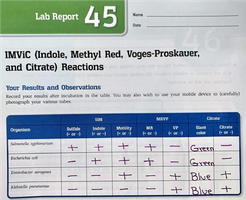
Positive result: Red color (stable acid production)
Negative result: Yellow color
Voges-Proskauer Test:
Reagents: VP-A (alpha-naphthol), VP-B (potassium hydroxide)
Positive result: Red color (acetoin present)
Negative result: No color change or copper color
Substrate: Glucose

Citrate Utilization Test
Principle and Interpretation
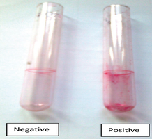
The citrate test determines the ability of an organism to use citrate as its sole carbon source. The medium contains bromthymol blue as a pH indicator.
Positive Result: Blue color (alkaline reaction, citrate utilized)
Negative Result: Green color (no citrate utilization)
Key Enzyme: Citrate lyase (citrase)
Substrate: Citrate
SIM Medium (Indole and Sulfur Reduction Tests)
Principle and Interpretation
SIM medium tests for sulfur reduction, indole production, and motility. Indole production is detected by adding Kovac's reagent after incubation.
Indole Test:
Substrate: Tryptophan
Enzyme: Tryptophanase
Positive result: Red ring after adding Kovac's reagent
Sulfur Reduction:
Substrate: Cysteine
Enzyme: Cysteine desulfurase
Positive result: Black precipitate (H2S production)
Catalase Test
Principle and Interpretation
The catalase test identifies organisms that produce the enzyme catalase, which breaks down hydrogen peroxide into water and oxygen.
Reagent: Hydrogen peroxide
Positive Result: Immediate bubbling (oxygen release)
Negative Result: No bubbling
Oxidase Test
Principle and Interpretation
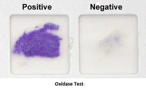
The oxidase test detects the presence of cytochrome c oxidase in bacteria. The reagent turns purple if the enzyme is present.
Reagent: Oxidase reagent (tetramethyl-p-phenylenediamine)
Positive Result: Purple color within 10-30 seconds
Negative Result: No color change or remains colorless
Nitrate Reduction Test
Principle and Interpretation
This test determines the ability of bacteria to reduce nitrate to nitrite or further to nitrogen gas or ammonia. It involves the addition of nitrate reagents A and B, and possibly zinc powder if no color change occurs.
Reagents: Nitrate reagent A (alpha-naphthylamine), Nitrate reagent B (sulfanilic acid), Zinc powder
Positive Result: Red color after reagents A and B (nitrite present), or no color after zinc (complete reduction)
Negative Result: Red color after zinc (nitrate not reduced)
Litmus Milk Medium
Principle and Interpretation
Litmus milk medium is used to differentiate bacteria based on their ability to ferment lactose, reduce litmus, and digest casein. The medium contains lactose, casein, and litmus as a pH and redox indicator.
Acid Reaction: Pink color (lactose fermentation)
Alkaline Reaction: Blue/purple color (casein digestion)
Curd Formation: Solidification due to acid or rennet production
Peptonization: Clearing of medium (complete proteolysis)
Starch Hydrolysis Test
Principle and Interpretation
This test identifies bacteria that produce amylase, which hydrolyzes starch into maltose. After incubation, iodine is added to detect the presence of starch.
Positive Result: Clear zone around growth after iodine addition (starch hydrolyzed)
Negative Result: Blue-black color (starch present)
Casein Hydrolysis Test
Principle and Interpretation
This test detects the ability of bacteria to hydrolyze casein, the major protein in milk, using the enzyme caseinase. A clear zone around the colony indicates casein hydrolysis.
Positive Result: Clear zone around growth (casein hydrolyzed)
Negative Result: No clearing (casein not hydrolyzed)
Gelatin Hydrolysis Test
Principle and Interpretation
This test determines the ability of bacteria to produce gelatinase, which hydrolyzes gelatin into amino acids. Liquefaction of the medium after refrigeration indicates a positive result.
Positive Result: Liquefied medium after refrigeration
Negative Result: Solid medium after refrigeration
Urea Hydrolysis Test
Principle and Interpretation
The urea hydrolysis test detects the production of urease, which hydrolyzes urea to ammonia and carbon dioxide, raising the pH and changing the color of the phenol red indicator.
Positive Result: Pink color (alkaline, ammonia produced)
Negative Result: No color change or yellow/orange
Effect of Temperature on Microbial Growth
Microbial Growth Ranges
Microorganisms are classified based on their optimal temperature ranges for growth:
Psychrophiles: Grow best at 0–15°C
Psychrotrophs: Grow at 0–30°C, optimal at 20–30°C
Mesophiles: Grow best at 25–40°C (human pathogens)
Thermophiles: Grow best at 50–60°C
Extreme Thermophiles: Grow at 80°C or higher
Fluid Thioglycolate Medium
Principle and Interpretation
This medium is used to determine the oxygen requirements of microorganisms. It contains thioglycolate, which reduces oxygen, creating an oxygen gradient.
Aerobes: Grow at the top
Anaerobes: Grow at the bottom
Facultative Anaerobes: Grow throughout the medium
Standard Plate Count
Principle and Interpretation
The standard plate count is used to estimate the number of viable bacteria in a sample. Plates with 30–300 colonies are considered countable. TFTC (too few to count) and TNTC (too numerous to count) are used for plates outside this range.
Application: Used in water, food, and clinical microbiology to assess contamination levels.
Antimicrobial Susceptibility Test (Kirby-Bauer Method)
Principle and Interpretation
This test evaluates the effectiveness of antibiotics against bacteria. Disks containing antibiotics are placed on an agar plate inoculated with the test organism. Zones of inhibition are measured to determine susceptibility.
Bactericidal: Kills bacteria
Bacteriostatic: Inhibits bacterial growth
ELISA (Enzyme-Linked Immunosorbent Assay)
Principle and Interpretation
ELISA is an immunological assay used to detect the presence of antigens or antibodies in a sample. There are direct and indirect ELISA types. Direct ELISA detects antigens, while indirect ELISA detects antibodies.
Applications: Diagnosis of diseases (HIV, Lyme disease), pregnancy tests, drug detection
Positive Result: Color change in the microtiter plate well
Bacterial Conjugation
Principle and Interpretation
Bacterial conjugation is a process of horizontal gene transfer where genetic material (usually plasmids) is transferred from one bacterium to another via direct contact, often through a pilus.
Significance: Contributes to genetic diversity and antibiotic resistance spread
Disadvantage: Can transfer undesirable traits, such as antibiotic resistance
Latex Agglutination Test
Principle and Interpretation
Latex agglutination is a rapid serological test used to detect the presence of antigens or antibodies. Latex beads coated with antibodies or antigens clump together in the presence of their specific target.
Positive Result: Visible clumping (agglutination)
Negative Result: No clumping
Applications: Diagnosis of diseases such as bacterial meningitis
