 Indietro
Microscopy and Laboratory Culture of Microorganisms: Core Concepts and Techniques
Guida di studio - Note intelligenti
 Appunti personalizzati basati sui tuoi materiali, ampliati con definizioni chiave, esempi e contesto.
Microscopy in Microbiology
Observing Microbes: Magnification and Resolution
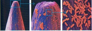
Microscopy is essential for visualizing microorganisms, which are too small to be seen with the naked eye. The two key properties of microscopes are magnification (enlarging the image) and resolution (the ability to distinguish two adjacent objects as separate entities).
Resolution is limited by the wavelength of light or electrons used. The human eye has a resolution of about 100 μm, while the best light microscopes achieve ~0.2 μm.
Magnification alone is not sufficient; enhanced resolution is necessary to see fine details.
Compound Light Microscope: Structure and Function
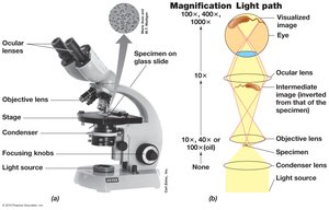
The compound light microscope is the most common tool for observing microbes. It uses visible light and two sets of lenses (objective and ocular) to magnify specimens.
Condenser focuses light onto the specimen.
Total magnification is calculated as:
The upper practical limit is about 2000X.
Improving Resolution: The Role of Immersion Oil
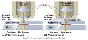
Resolution depends on the wavelength of light and the numerical aperture of the lens. Immersion oil is used to increase the amount of light entering the objective lens, thereby improving resolution.
Without oil, light is refracted and lost; with oil, more light is captured by the lens.
Best resolution with a light microscope is about 0.2 μm.
Types of Light Microscopy
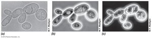
Bright-field microscopy: Relies on differences in contrast; often requires staining to visualize bacteria.
Phase-contrast microscopy: Enhances contrast by amplifying differences in refractive index; useful for viewing live, unstained cells.
Dark-field microscopy: Only scattered light reaches the lens, making cells appear bright against a dark background.
Staining Techniques
Staining increases contrast in bright-field microscopy. Most stains are basic dyes that bind to negatively charged cell components.
Common stains: methylene blue, crystal violet, safranin.
Staining procedure: smear preparation, heat fixing, staining, and microscopy.
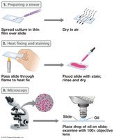
Differential Stains: The Gram Stain
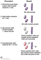
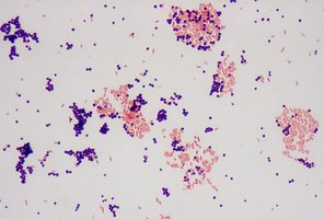
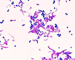
The Gram stain differentiates bacteria based on cell wall structure, dividing them into Gram-positive (purple) and Gram-negative (pink) groups.
Gram-positive bacteria retain crystal violet-iodine complex after decolorization.
Gram-negative bacteria are decolorized and take up the counterstain (safranin).
Acid-Fast Stain
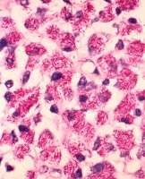
The acid-fast stain is used to identify bacteria with waxy, mycolic acid-rich cell walls, such as Mycobacterium species.
Acid-fast cells retain the red primary stain; non-acid-fast cells take up the blue counterstain.
Fluorescence Microscopy
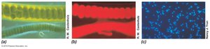
Fluorescence microscopy uses dyes or naturally fluorescent molecules to visualize cells. Fluorescent dyes absorb light at one wavelength and emit it at another, allowing for specific labeling of cell components.
Example: DAPI binds DNA and emits blue fluorescence.
Useful for identifying specific microbes in mixed samples.
Differential Interference Contrast (DIC) Microscopy
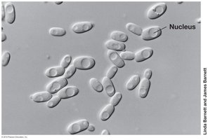
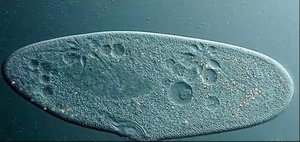
DIC microscopy uses polarized light and prisms to enhance contrast and produce a pseudo-3D effect, making internal structures like nuclei and vacuoles more visible.
Especially useful for observing eukaryotic microbes and cellular inclusions.
Confocal Scanning Laser Microscopy
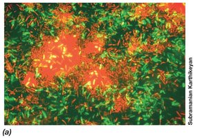
Confocal microscopy uses lasers and computer processing to generate high-resolution, three-dimensional images of specimens, such as microbial biofilms. It allows for optical sectioning and reconstruction of thick samples.
Resolution can reach 0.1 μm.
Enables visualization of cells at different depths within a biofilm.
Electron Microscopy
Electron microscopes use electron beams instead of light, providing much higher resolution due to the shorter wavelength of electrons. Two main types are:
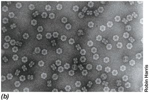
Transmission Electron Microscope (TEM): Electrons pass through ultrathin sections of specimens, revealing internal structures. Resolution is about 0.2 nm.
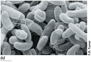
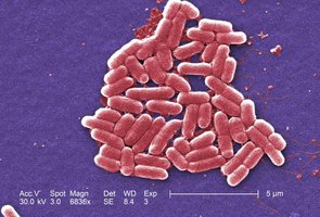
Scanning Electron Microscope (SEM): Electrons scan the specimen surface, producing detailed 3D images of external features.
Laboratory Culture of Microorganisms
Essential Nutrients for Microbial Growth
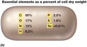
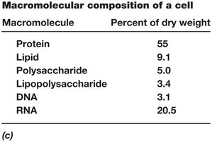
Microbes require a variety of nutrients for growth, which must be supplied in laboratory culture media. These include macronutrients (needed in large amounts) and micronutrients (trace elements).
Macronutrients: C, N, O, H, P, S
Micronutrients: Se, and others, typically <0.01% of cell mass
Types of Culture Media
Culture media provide the nutrients required for microbial growth. They can be classified as:
Defined media: Exact chemical composition is known.
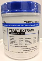
Complex media: Contain extracts or digests of natural products; composition is not precisely known (e.g., yeast extract, tryptic soy broth).
Specialized Media: Selective, Differential, and Enriched
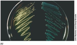
Selective media: Inhibit growth of some microbes while allowing others to grow (e.g., eosin-methylene blue agar inhibits Gram-positive bacteria).
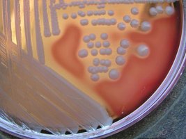
Differential media: Allow distinction between different microbes based on colony appearance or color changes (e.g., lactose fermenters on EMB agar appear with a green metallic sheen).
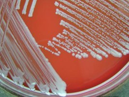
Enriched media: Supplemented with additional nutrients for fastidious organisms (e.g., blood agar, chocolate agar).
Preparation and Sterilization of Culture Media
Culture media must be sterilized, typically by autoclaving, to prevent contamination. Agar is often added to solidify media, allowing for the growth of isolated colonies.
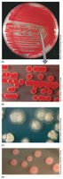
Solid media enable the formation of colonies, which can vary in shape, size, and color depending on the organism and conditions.
Aseptic Technique
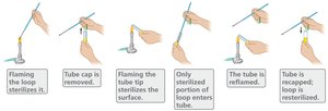
Aseptic technique is critical to prevent contamination of cultures and sterile media during handling. This involves sterilizing instruments and careful transfer of samples.
Flaming loops and tube openings are common steps.
Isolation of Pure Cultures: Streak Plate and Spread Plate Methods
Pure cultures are obtained by isolating single colonies on solid media. The two main methods are:
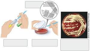
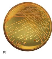
Streak plate: Involves spreading a loopful of culture over the surface of an agar plate in a pattern that dilutes the sample, resulting in isolated colonies.
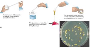
Spread plate: Involves spreading a diluted sample evenly over the surface of an agar plate using a sterile spreader.
Summary Table: Types of Culture Media
Type of Media | Description | Example |
|---|---|---|
Defined | Exact chemical composition known | Minimal salts medium |
Complex | Contains digests/extracts; composition not precisely known | Yeast extract broth |
Selective | Inhibits some microbes, allows others | EMB agar |
Differential | Distinguishes microbes by colony appearance or color change | Blood agar |
Enriched | Supplemented for fastidious organisms | Chocolate agar |
