 Indietro
Microscopy, Staining, and Classification in Microbiology
Guida di studio - Note intelligenti
 Appunti personalizzati basati sui tuoi materiali, ampliati con definizioni chiave, esempi e contesto.
Microscopy, Staining, and Classification
Introduction
This chapter explores the foundational principles and techniques of microscopy, staining, and the classification of microorganisms. Understanding these concepts is essential for visualizing, identifying, and categorizing microbes in the laboratory and clinical settings.
General Principles of Microscopy
Wavelength of Radiation
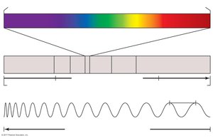
The wavelength of radiation used in microscopy determines the resolving power of the microscope. Shorter wavelengths provide higher resolution, allowing for the visualization of smaller structures.
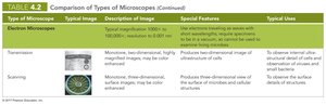
Visible light is commonly used in light microscopy, but electron microscopes use much shorter wavelengths (electrons) for greater resolution.
Resolution is limited by the wavelength; objects smaller than half the wavelength cannot be resolved.
Magnification
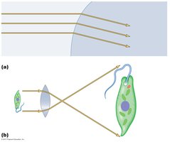
Magnification is the process of enlarging the appearance of an object. It is achieved by using lenses to bend light and form an image that is larger than the actual specimen.
Magnification is calculated as the product of the magnifications of the objective and ocular lenses.
Convex lenses are used to focus light and create an enlarged, inverted image.
Resolution
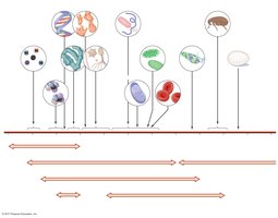
Resolution is the ability to distinguish two points as separate entities. It is a critical factor in microscopy, as higher resolution allows for clearer and more detailed images of microorganisms.
Resolution depends on the wavelength of light and the numerical aperture of the lens.
Electron microscopes provide much higher resolution than light microscopes.
Contrast
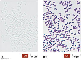
Contrast refers to the differences in intensity between an object and its background. High contrast is essential for distinguishing structures within a specimen.
Staining techniques are often used to increase contrast.
Phase-contrast and differential interference contrast microscopes enhance contrast without staining.
Types of Microscopes
Light Microscopy
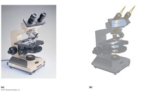
Light microscopes use visible light to illuminate specimens. There are several types, each with unique features and applications.
Bright-field microscopes: The most common type; specimens appear dark against a bright background.
Simple microscopes: Contain a single lens (e.g., Leeuwenhoek's microscope).
Compound microscopes: Use a series of lenses for greater magnification and resolution.
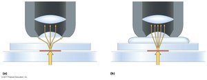
Oil immersion: Increases resolution by reducing light refraction.
Comparison of Types of Microscopes
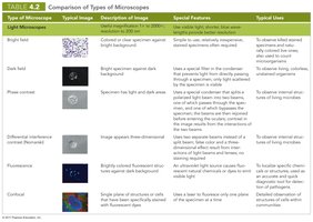
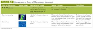
The following tables summarize the main types of microscopes, their features, and typical uses.
Type of Microscope | Typical Image | Description of Image | Special Features | Typical Uses |
|---|---|---|---|---|
Bright field | Colored or clear specimen against bright background | Simple to use; requires staining | Routine observation of stained specimens | General laboratory use |
Dark field | Bright specimen against dark background | Enhances contrast in unstained samples | Observation of living, unstained organisms | Motility studies |
Phase contrast | Specimen appears with varying degrees of brightness and contrast | Enhances contrast without staining | Observation of internal structures in living cells | Cellular detail studies |
Differential interference contrast (Nomarski) | 3D-like images | Uses two beams of light | Detailed observation of cell structures | Cell morphology |
Fluorescence | Brightly colored fluorescent structures | Uses UV light and fluorescent dyes | Identification of specific microbes | Immunofluorescence |
Confocal | Single plane of structures or cells | Uses lasers for optical sectioning | 3D reconstruction of specimens | Biofilm studies |
Staining Techniques
Principles of Staining
Staining enhances contrast and resolution by coloring microorganisms or their structures. Dyes are typically salts with a colored ion (chromophore).
Acidic dyes stain alkaline structures; basic dyes stain acidic structures (most cells are negatively charged, so basic dyes are more common).
Staining is essential for observing cell morphology, arrangement, and internal structures.
Simple Stains
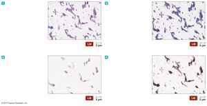
Simple stains use a single basic dye to color cells, making it easier to observe their size, shape, and arrangement.
Examples: Crystal violet, safranin, methylene blue.
Differential Stains
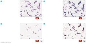
Differential stains use more than one dye to distinguish between different types of cells or structures.
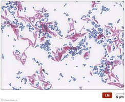
Gram stain: Differentiates Gram-positive (purple) and Gram-negative (pink) bacteria.
Acid-fast stain: Identifies Mycobacterium and Nocardia species.
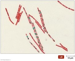
Endospore stain: Highlights endospores in genera Bacillus and Clostridium.
Histological stains: Used for tissue samples.
Special Stains
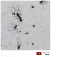
Special stains are used to highlight specific microbial structures, such as capsules, flagella, or endospores.
Negative stains: Stain the background, leaving cells colorless.
Flagellar stains: Make bacterial flagella visible.
Fluorescent stains: Bind to specific cell components and fluoresce under UV light.
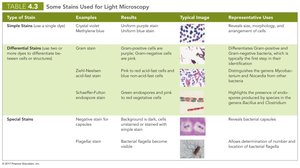
Summary Table: Some Stains Used for Light Microscopy
Type of Stain | Examples | Typical Image | Representative Uses |
|---|---|---|---|
Simple Stains | Crystal violet, methylene blue | Uniform purple or blue stain | Reveals cell morphology and arrangement |
Differential Stains | Gram, Ziehl-Neelsen, Schaeffer-Fulton | Multiple colors (e.g., purple/blue and pink/red) | Distinguishes cell types, highlights endospores |
Special Stains | Negative, flagellar | Background or flagella stained | Reveals capsules, flagella |
Classification and Identification of Microorganisms
Taxonomy: Classification, Nomenclature, and Identification
Taxonomy is the science of classifying organisms. It involves organizing, naming, and identifying organisms based on shared characteristics and evolutionary relationships.
Linnaeus developed the binomial nomenclature system and classified organisms into kingdoms.
Modern taxonomy reflects phylogenetic relationships, often based on genetic analysis (e.g., rRNA sequences).
Carl Woese proposed three domains: Bacteria, Archaea, and Eukarya.
Taxonomic and Identifying Characteristics
Microorganisms are identified using a combination of physical, biochemical, serological, and genetic characteristics.
Physical characteristics: Morphology, colony appearance.
Biochemical tests: Ability to utilize or produce specific chemicals.
Serological tests: Antigen-antibody reactions.
Phage typing: Susceptibility to bacteriophages.
Analysis of nucleic acids: Genetic sequencing.
Dichotomous Keys
Dichotomous keys are tools used to identify organisms through a series of paired statements, guiding the user to the correct identification based on observable traits.
Each step presents two choices; the correct choice leads to the next step or the identification of the organism.
Commonly used in microbiology for bacterial identification.
Summary
Microscopy, staining, and classification are essential techniques in microbiology for the visualization, differentiation, and identification of microorganisms. Mastery of these concepts enables accurate laboratory analysis and enhances our understanding of microbial diversity and relationships.
