 Indietro
MMBIO 221 Exam 1 Review: Foundations of Microbiology
Guida di studio - Note intelligenti
 Appunti personalizzati basati sui tuoi materiali, ampliati con definizioni chiave, esempi e contesto.
Major Pioneers in Microbiology
Historical Figures and Their Contributions
The field of microbiology was shaped by numerous scientists whose discoveries laid the foundation for modern understanding of microbes and infectious disease.
Aristotle: Proposed spontaneous generation, the idea that life could arise from nonliving matter.
Francesco Redi: Disproved spontaneous generation with meat and flask experiments.
Antoni van Leeuwenhoek: Father of microscopy; first to observe microorganisms, which he called "animalcules."
Carolus Linnaeus: Developed binomial nomenclature and modern taxonomy.
Lazzaro Spallanzani: Further disproved spontaneous generation using sealed broth flasks.
Edward Jenner: Developed the first smallpox vaccine using cowpox virus.
Ignaz Semmelweis: Demonstrated that handwashing prevents infection.
John Snow: Father of epidemiology; stopped a cholera outbreak by removing a contaminated water pump.
Louis Pasteur: Disproved spontaneous generation with swan-neck flasks, invented pasteurization, and developed vaccines for rabies, fowl cholera, and anthrax.
Joseph Lister: Pioneered aseptic surgery using phenol.
Robert Koch: Developed Koch’s postulates, Petri plates, and staining techniques; established causative links between microbes and disease.
Christian Gram: Developed the Gram stain, a key differential staining technique.
Paul Ehrlich: Sought a "magic bullet" for pathogens; discovered Salvarsan for syphilis.
Alexander Fleming: Discovered penicillin, the first antibiotic.
Selman Waksman: Discovered streptomycin and coined the term "antibiotic."
Robert Whittaker: Proposed the five-kingdom classification system.
Carl Woese: Proposed the three-domain system (Bacteria, Archaea, Eukarya) based on rRNA sequencing.
Classification and Characteristics of Microbes
Major Groups of Microorganisms
Microbes are classified based on cellular structure, mode of nutrition, and other characteristics.
Fungi: Eukaryotic, absorb nutrients, cell walls of chitin, classified by sexual spores. Includes unicellular yeasts and multicellular molds.
Algae: Eukaryotic, photosynthetic, can be unicellular or multicellular, classified by pigments and cell wall composition.
Protozoa: Eukaryotic, unicellular, ingest nutrients, classified by motility mechanisms.
Bacteria: Prokaryotic, reproduce by binary fission, classified by cell wall structure, shape, and oxygen requirements.
Viruses: Obligate intracellular parasites, not cellular, covered in later chapters.
Prokaryotic vs. Eukaryotic Cells
Key Differences
Prokaryotic and eukaryotic cells differ in complexity, size, and internal organization.
Prokaryotes: No nucleus, smaller (∼1 μm), simpler structure.
Eukaryotes: True nucleus, larger (10–100 μm), more complex.

Endosymbiotic Theory
Origin of Eukaryotic Organelles
The endosymbiotic theory explains the origin of mitochondria and chloroplasts as formerly free-living prokaryotes engulfed by ancestral eukaryotic cells.
Mitochondria and chloroplasts have their own DNA and double membranes, supporting this theory.
Structure and Function of Prokaryotic Cell Components
External and Internal Structures
Axial Filaments: Internal flagella in spirochetes for corkscrew motility.
Cell Wall: Peptidoglycan structure for shape and osmotic protection.
Cell Membrane: Phospholipid bilayer for selective transport and metabolism.
Cytoplasm: Gel-like matrix for metabolic reactions.
Capsule: Polysaccharide layer for protection and adherence.
Endospore: Dormant, resistant structure for survival in harsh conditions.
Fimbriae: Attachment structures for surfaces and biofilms.
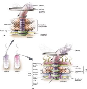
Flagella: Motility appendages; composed of filament, hook, and basal body.
Glycocalyx: Sticky layer for attachment and protection.
Nucleoid: Region with circular DNA chromosome.
Plasmid: Small, extrachromosomal DNA with accessory genes.
Pili: Attachment and DNA transfer structures.
Ribosome: Site of protein synthesis (70S in prokaryotes).
Flagella Structure and Function
Composed of filament, hook, and basal body.
Movement: "Runs" (toward stimuli) and "tumbles" (away from stimuli).
Cell Membranes and Transport Processes
Membrane Structure and Permeability
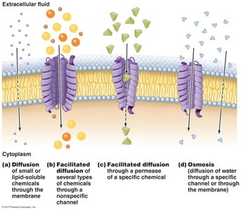
The cell membrane is a selectively permeable phospholipid bilayer. Transport processes include passive and active mechanisms.
Passive Transport: No energy required.
Simple diffusion: Small, nonpolar molecules move down concentration gradient.
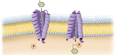
Facilitated diffusion: Larger or charged molecules move via transport proteins.
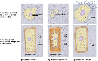
Osmosis: Water movement to balance solute concentrations.
Osmosis and Tonicity
Hypertonic: Cell shrinks (crenation/plasmolysis).
Hypotonic: Cell swells; may burst if no cell wall.
Isotonic: No net water movement; cell remains normal.
Active Transport
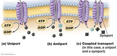
Requires energy (ATP).
Types: Uniport (one substance), symport (two substances same direction), antiport (opposite directions), group translocation (chemical modification during transport).
Gram-Positive, Gram-Negative, and Acid-Fast Bacteria
Unique and Common Features
Gram-Positive: Thick peptidoglycan, teichoic acids.
Gram-Negative: Outer membrane, thin peptidoglycan, lipopolysaccharide (LPS), Lipid A (endotoxin).
Acid-Fast: Mycolic acids, arabinogalactan, high lipid content.
Common: Cell membrane, cytoplasm, nucleoid, ribosomes, cell wall (structurally different), flagella, pili/fimbriae, plasmids.
Bacteria Lacking a Cell Wall
Mycoplasma: Bacterial genus lacking a cell wall; resistant to antibiotics targeting cell wall synthesis.
Principles of Microscopy
Magnification, Resolution, Contrast, and Wavelength
Magnification: Enlarges image of specimen.
Resolution: Ability to distinguish two close objects.
Contrast: Difference between specimen and background; enhanced by staining.
Wavelength: Determines resolving power; shorter wavelengths (e.g., electron beams) resolve smaller structures.
Types of Microscopy
Bright-Field: Stained specimens; cell shape and arrangement.
Dark-Field: Live, thin organisms; bright specimen on dark background.
Phase-Contrast: Live, unstained cells; internal structures.
Differential Interference Contrast: 3D-like imaging of live cells.
Fluorescence: Specific detection using fluorescent dyes/antibodies.
Confocal: 3D imaging of thick samples using lasers.
Transmission Electron Microscopy (TEM): Internal ultrastructure; nanometer resolution.
Scanning Electron Microscopy (SEM): Surface morphology in 3D.
Staining Techniques
Gram Stain Procedure
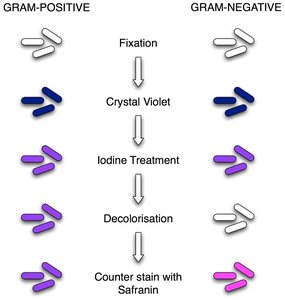
Crystal violet (primary stain): All cells purple.
Iodine (mordant): Fixes stain; all cells purple.
Decolorizer (alcohol/acetone): Gram-positive remain purple; Gram-negative lose color.
Safranin (counterstain): Gram-positive stay purple; Gram-negative turn pink.
Other Staining Methods
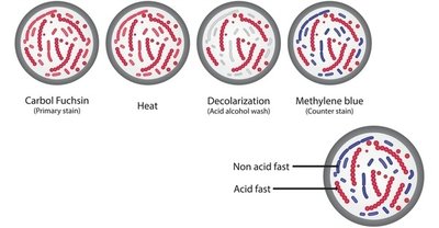
Acid-Fast Stain: Acid-fast cells are fuchsia (red); non-acid-fast are blue.
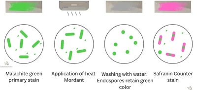
Endospore Stain: Endospores are green; vegetative cells are pink ("Christmas colors").
Enzymes and Metabolism
Structure and Function of Enzymes
Enzymes are biological catalysts, mostly proteins (some RNA ribozymes).
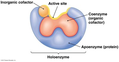
Apoenzyme: Protein portion without cofactor.
Cofactor: Non-protein helper (inorganic or organic/coenzyme).
Holoenzyme: Complete, active enzyme with all cofactors bound.
Active Site: Region where substrate binds.
Factors Affecting Enzyme Activity
Substrate concentration (increases rate to a point).
Temperature (increases rate to optimum, then denaturation).
pH (extremes denature enzymes).
Ionic strength (each enzyme has an optimum).
Inhibitors: Competitive (bind active site), non-competitive (allosteric site), feedback inhibition (product inhibits enzyme).
Enzymatic Reactions and Oxygen Detoxification
Enzymes act on substrates to catalyze reactions.
Key enzymes for detoxifying oxygen radicals:
Catalase:
Peroxidase:
Superoxide dismutase:
Microbial Nutrition and Growth
Types of Culture Media
Selective: Inhibits some microbes, allows others.
Differential: Distinguishes microbes by biochemical reactions (color changes).
Defined: Exact chemical composition known.
Enriched: Extra nutrients for fastidious organisms.
Complex: Contains ingredients of unknown composition (e.g., yeast extract).
Basic Growth Requirements and Nutritional Types
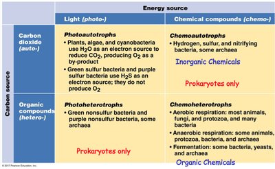
Organisms classified by carbon and energy sources:
Energy Source | Carbon Source | Example |
|---|---|---|
Light (photo-) | CO2 (auto-) | Photoautotrophs: Plants, algae, cyanobacteria |
Chemicals (chemo-) | CO2 (auto-) | Chemoautotrophs: Nitrifying bacteria |
Light (photo-) | Organic compounds (hetero-) | Photoheterotrophs: Purple non-sulfur bacteria |
Chemicals (chemo-) | Organic compounds (hetero-) | Chemoheterotrophs: Most animals, fungi, protozoa, many bacteria |
Environmental Effects on Microbial Growth
Temperature: Psychrophiles (cold), mesophiles (moderate), thermophiles (hot), hyperthermophiles (very hot).
pH: Neutrophiles (neutral), acidophiles (acidic), alkaliphiles (basic).
Osmotic Pressure: Halophiles (high salt).
Pressure: Barophiles (high pressure).
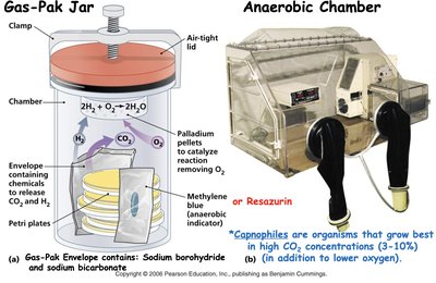
CO2: Capnophiles (high CO2).
Anaerobic Culturing and Gas-Pak System
Gas-Pak releases CO2 and H2 to remove O2 and create anaerobic conditions for obligate anaerobes.
Microbial Growth Phases
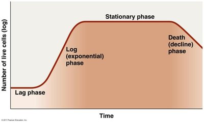
Batch Culture Growth Curve
Lag Phase: Cells adapt to environment, synthesize enzymes.
Log (Exponential) Phase: Rapid cell division and growth.
Stationary Phase: Growth rate equals death rate; nutrients deplete.
Death Phase: Cells die due to lack of nutrients and waste accumulation.
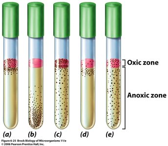
Oxygen Requirements and Thioglycolate Medium
Obligate Aerobe: Grows only at top (oxic zone).
Obligate Anaerobe: Grows only at bottom (anoxic zone).
Facultative Anaerobe: Grows throughout, best at top.
Microaerophile: Grows just below surface.
Aerotolerant Anaerobe: Grows evenly throughout.
Metabolic Concepts
Anaerobic Respiration
Uses terminal electron acceptors other than oxygen (e.g., nitrate, sulfate).
Inhibitors vs. Uncouplers of Oxidative Phosphorylation
Inhibitors: Block electron transport, halt ATP synthesis and O2 consumption (e.g., cyanide).
Uncouplers: Allow electron flow but collapse proton gradient, reducing ATP synthesis and releasing energy as heat (e.g., dinitrophenol).
ATP Synthesis
Number of protons required to synthesize one cytoplasmic ATP in eukaryotes: 4.
NAD and FAD
NAD (Nicotinamide adenine dinucleotide) and FAD (Flavin adenine dinucleotide) are dinucleotide cofactors that carry electrons and hydrogen during metabolic reactions.
Biological Macromolecules
Subunits and Examples
Proteins: Amino acids (e.g., enzymes, antibodies).
Lipids: Glycerol and fatty acids (e.g., phospholipids, steroids).
Carbohydrates: Monosaccharides (e.g., cellulose, glycogen).
Nucleic Acids: Nucleotides (e.g., DNA, RNA).
