Indietro
Observing Microorganisms Through a Microscope
Guida di studio - Note intelligenti
 Appunti personalizzati basati sui tuoi materiali, ampliati con definizioni chiave, esempi e contesto.
Observing Microorganisms Through a Microscope
Introduction to Microscopy
Microscopy is essential in microbiology for visualizing organisms too small to be seen with the naked eye. The development of microscopes has enabled scientists to study the structure, function, and classification of microorganisms.
Size, Resolution, and Types of Microscopes
Size and Resolution
Resolution is the ability of a lens to distinguish two points as separate entities. The resolving power of a microscope is determined by the wavelength of light used; shorter wavelengths provide greater resolution. For example, a microscope with a resolving power of 0.4 nm can distinguish between two points that are at least 0.4 nm apart.
Simple microscope: Contains a single lens.
Compound microscope: Uses multiple lenses to achieve higher magnification and resolution.
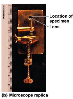
Compound Light Microscope
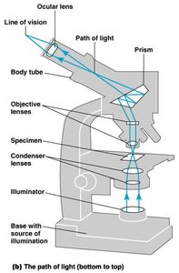
The compound light microscope is the most commonly used instrument in microbiology. It uses a series of lenses to magnify the image of a specimen. The total magnification is the product of the magnification of the objective lens and the ocular lens.
Objective lens: Closest to the specimen; provides initial magnification.
Ocular lens: Further magnifies the image formed by the objective lens.
Total magnification:
Refractive Index and Immersion Oil
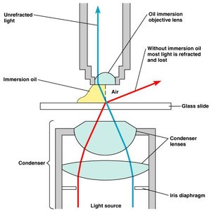
The refractive index is the measure of how much a medium bends light. When using high-magnification lenses, light may bend so much in air that it misses the lens. Immersion oil is used to reduce light refraction and increase resolution by matching the refractive index of glass.
Types of Light Microscopy
Brightfield and Darkfield Illumination
Different illumination techniques enhance contrast and detail in specimens:
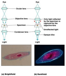
Brightfield microscopy: Stained specimens appear dark against a bright background. Most commonly used for fixed, stained samples.
Darkfield microscopy: Unstained specimens appear bright against a dark background. Useful for observing live, delicate, or hard-to-stain organisms.
Phase-Contrast Microscopy
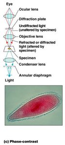
Phase-contrast microscopy accentuates differences in the refractive index of cellular components, allowing visualization of internal structures in living cells without staining or fixing.
Light waves passing through the specimen are diffracted and interfere with each other, enhancing contrast.
Differential Interference Contrast (DIC) Microscopy
DIC microscopy uses two beams of light and accentuates differences in refractive index, producing high-contrast, pseudo-3D images of living cells.
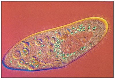
Fluorescence and Confocal Microscopy
Fluorescence microscopy uses ultraviolet (UV) light to excite fluorescent dyes (fluorochromes) that emit visible light. It is widely used for detecting specific microbes or cellular components, often with fluorescent-antibody techniques (immunofluorescence).
Fluorescent substances absorb UV light and emit visible light.
Immunofluorescence uses antibodies labeled with fluorochromes to identify pathogens.
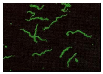
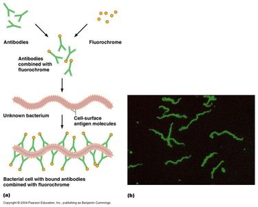
Confocal microscopy uses lasers and fluorochromes to scan specimens and produce high-resolution, three-dimensional images by focusing on a single plane at a time.
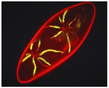
Electron Microscopy
Principles and Types
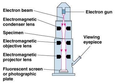
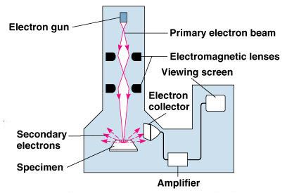
Electron microscopes use beams of electrons instead of light, providing much greater resolution (up to 2.5 nm for TEM). There are two main types:
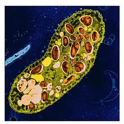
Transmission Electron Microscopy (TEM): Electrons pass through ultrathin sections of specimens, revealing internal structures. Specimens are often stained with heavy metals.
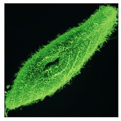
Scanning Electron Microscopy (SEM): Electrons scan the surface of a specimen, and secondary electrons emitted from the surface are detected to produce detailed images of surface structures.
Preparation and Staining of Specimens
Preparation for Light Microscopy
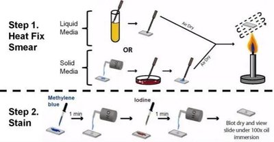
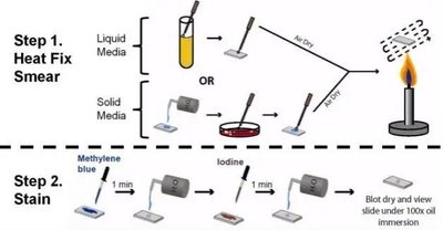
Microbial specimens are typically prepared as a smear on a glass slide, which is then fixed (by heat or chemicals) to attach and kill the microbes. Staining increases contrast and allows for better visualization of cellular structures.
Types of Stains
Simple stains: Use a single basic dye to color cells, making them easier to see.
Differential stains: Distinguish between different types of bacteria or structures. Examples include the Gram stain and acid-fast stain.
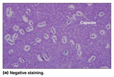
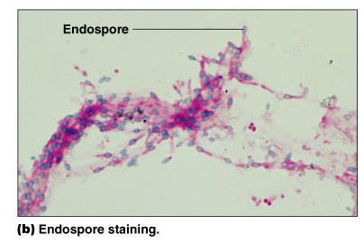
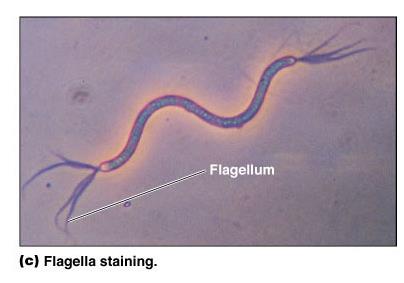
Special stains: Used for specific structures such as capsules, endospores, and flagella.
Gram Stain
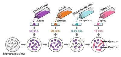
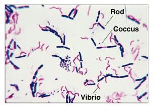
The Gram stain is a differential staining technique that classifies bacteria as Gram-positive or Gram-negative based on differences in cell wall structure. The procedure involves four steps: application of crystal violet (primary stain), iodine (mordant), alcohol-acetone (decolorizer), and safranin (counterstain).
Step | Gram + Color | Gram - Color |
|---|---|---|
Crystal violet | Purple | Purple |
Iodine | Purple | Purple |
Alcohol-acetone | Purple | Colorless |
Safranin | Purple | Red |
Acid-Fast Stain
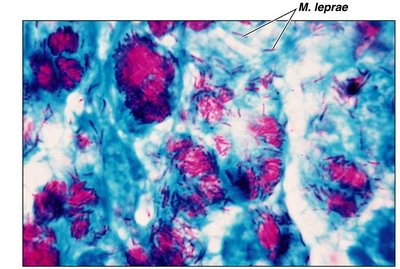
The acid-fast stain identifies bacteria with waxy cell walls (e.g., Mycobacterium species). Acid-fast cells retain the primary stain (carbolfuchsin) even after decolorization with acid-alcohol, while non-acid-fast cells do not.
Special Stains
Negative staining: Stains the background, leaving capsules visible as clear halos.
Endospore staining: Uses heat to drive the stain into resistant endospores.
Flagella staining: Uses a mordant to thicken flagella for visibility under the microscope.