 Indietro
Observing Microorganisms through a Microscope: Microscopy and Staining Techniques
Guida di studio - Note intelligenti
 Appunti personalizzati basati sui tuoi materiali, ampliati con definizioni chiave, esempi e contesto.
Observing Microorganisms through a Microscope
Units of Measurement
Microorganisms are measured using units much smaller than those used in everyday life. Understanding these units is essential for interpreting microscopic observations.
Micrometer (μm): One micrometer is 1/1,000,000 of a meter (10-6 m).
Nanometer (nm): One nanometer is 1/1,000,000,000 of a meter (10-9 m).
Conversion: 1 μm = 1,000 nm.
Application: Most bacteria are measured in micrometers, while viruses are measured in nanometers.
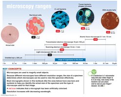
Example: If a bacterium is 10 μm long, it is 10,000 nm in length.
Microscopy: The Instruments
Microscopes are essential tools in microbiology, allowing visualization of organisms too small to be seen with the naked eye. There are several types of microscopes, each with unique features and applications.
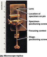
Simple Microscope: Contains a single lens, similar to the original designs by Anton van Leeuwenhoek.
Compound Microscope: Uses multiple lenses to magnify specimens, providing higher magnification and resolution.
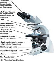
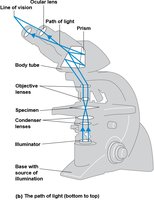
Path of Light in a Compound Microscope
Understanding the path of light is crucial for proper use and interpretation of compound microscopes.
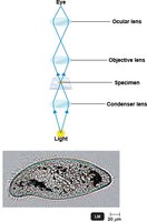
Light passes through the illuminator, condenser lenses, specimen, objective lenses, body tube, and finally the ocular lens.
Total Magnification: Calculated as the product of the magnification of the objective lens and the ocular lens.
Resolution: The ability to distinguish two points as separate; higher resolution allows for clearer images.
Shorter wavelengths of light provide greater resolution.
Refractive Index and Immersion Oil
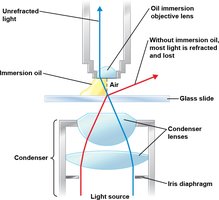
The refractive index is a measure of how much a medium bends light. Immersion oil is used to reduce light refraction and increase resolution when using high-power objective lenses.
Immersion oil: Prevents loss of light due to refraction, allowing more light to enter the objective lens.
Types of Light Microscopy
Light microscopes use visible light to observe specimens. Several specialized types exist, each suited for different applications.
Brightfield Microscopy: Dark objects are visible against a bright background; commonly used for stained specimens.
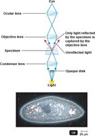
Darkfield Microscopy: Light objects are visible against a dark background; useful for observing live, unstained specimens.
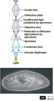
Phase-Contrast Microscopy: Enhances contrast of transparent specimens; allows examination of living cells and internal structures.
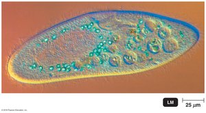
Differential Interference Contrast (DIC) Microscopy: Uses two light beams and prisms to provide high-contrast, colorful images.
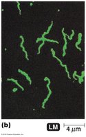
Fluorescence Microscopy: Uses UV light; specimens are stained with fluorescent dyes and emit visible light.
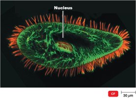
Confocal Microscopy: Uses fluorochrome dyes and laser light to produce three-dimensional images.
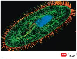
Two-Photon Microscopy: Uses two photons of long-wavelength light to study living cells deep within tissues.
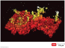
Scanning Acoustic Microscopy: Uses sound waves to study cells attached to surfaces.
Electron Microscopy
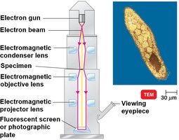
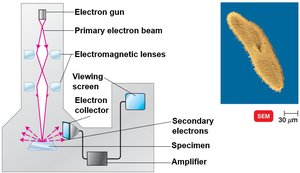
Electron microscopes use electron beams instead of light, allowing for much greater resolution and magnification. They are essential for visualizing viruses and detailed cell structures.
Transmission Electron Microscope (TEM): Electrons pass through thin sections of specimens, revealing internal structures.
Scanning Electron Microscope (SEM): Electrons scan the surface of specimens, producing three-dimensional images of surface structures.
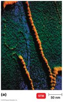
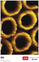
Scanned-Probe Microscopes: Use probes to scan surfaces at atomic resolution.
Preparation of Specimens for Light Microscopy
Proper specimen preparation is crucial for accurate microscopic observation. Staining techniques enhance contrast and allow for differentiation of cellular structures.
Staining: Coloring microorganisms with dyes to emphasize certain structures.
Smear: Thin film of material containing microorganisms spread over a slide.
Fixation: Attaches and kills microorganisms, preserving their structure.
Basic Dye: Chromophore is a cation; stains cells directly.
Acidic Dye: Chromophore is an anion; stains background (negative staining).
Simple Stains
Simple stains use a single basic dye to highlight the entire microorganism, making cell shapes and structures visible. A mordant may be used to enhance staining.
Purpose: Visualize cell morphology and arrangement.
Mordant: Substance that holds the stain or coats the specimen to enlarge it.
Differential Stains
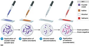
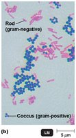
Differential stains distinguish between different types of bacteria. The most common are the Gram stain and the acid-fast stain.
Gram Stain: Classifies bacteria as gram-positive (thick peptidoglycan cell wall) or gram-negative (thin peptidoglycan cell wall and outer membrane).
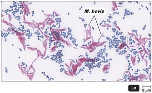
Acid-Fast Stain: Identifies bacteria with waxy cell walls, such as Mycobacterium and Nocardia.
Special Stains
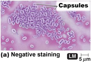
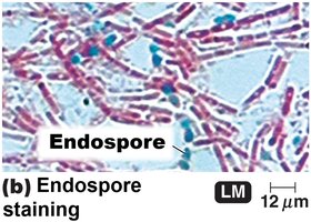
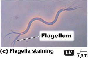
Special stains are used to highlight specific structures within microorganisms, such as capsules, endospores, and flagella.
Capsule Stain: Negative staining with India ink or nigrosin; capsules appear as halos around cells.
Endospore Stain: Uses malachite green and heat; endospores appear green within red or pink cells.
Flagella Stain: Uses a mordant and carbolfuchsin to visualize flagella.
Summary Table: Types of Microscopy and Their Applications
Microscopy Type | Principle | Application |
|---|---|---|
Brightfield | Visible light, stained specimens | General observation of cell morphology |
Darkfield | Light reflected by specimen | Live, unstained specimens |
Phase-Contrast | Combines direct and diffracted light | Internal cell structures, living cells |
DIC | Two beams, prisms | High-contrast, colorful images |
Fluorescence | UV light, fluorescent dyes | Identification, immunofluorescence |
Confocal | Laser, fluorochrome dyes | 3D imaging |
Two-Photon | Two photons, deep tissue | Live cell imaging |
Scanning Acoustic | Sound waves | Cells on surfaces |
TEM | Electron beam through specimen | Internal structures, viruses |
SEM | Electron beam scans surface | Surface structures, 3D images |
STM/AFM | Probe scans surface | Atomic resolution, surface details |
Additional info: This summary table is inferred from the context and standard microbiology textbooks.
