 Indietro
Prokaryotes: Structure, Classification, and Biofilms
Guida di studio - Note intelligenti
 Appunti personalizzati basati sui tuoi materiali, ampliati con definizioni chiave, esempi e contesto.
Prokaryotes: Structure, Classification, and Biofilms
Introduction to Prokaryotes
Prokaryotes are unicellular organisms that lack a membrane-bound nucleus and organelles. They are classified into two domains: Bacteria and Archaea. Understanding their structure, classification, and life cycles is fundamental to microbiology.
Classification of Life: Historical and Modern Perspectives
Five Kingdom and Three Domain Systems
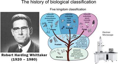
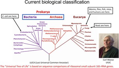
Biological classification has evolved from the five-kingdom system to the three-domain system based on molecular data, particularly 16S rRNA gene sequences.
Five Kingdoms: Monera, Protista, Fungi, Plantae, Animalia
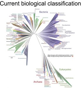
Three Domains: Bacteria, Archaea, Eukarya
16S rRNA gene sequencing is the molecular basis for modern classification, distinguishing the three domains.
Bacterial Cell Structure and Morphology
Key Structural Features
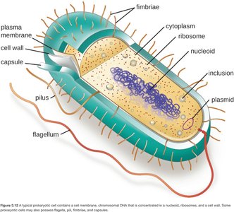
Bacterial cells possess unique structures that contribute to their survival and function. These include the cell wall, plasma membrane, capsule, pili, flagella, and genetic material.
Cell Wall: Provides shape and protection; composition distinguishes Gram-positive and Gram-negative bacteria.
Capsule: A polysaccharide layer that aids in protection and adherence.
Plasma Membrane: Regulates transport of substances in and out of the cell.
Pili and Fimbriae: Involved in attachment and conjugation.
Flagella: Enable motility.
Nucleoid: Region containing the bacterial chromosome.
Plasmids: Small, circular DNA molecules carrying accessory genes.
Bacterial Shapes and Arrangements
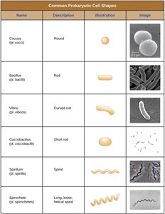
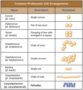
Bacteria exhibit a variety of shapes and arrangements, which are important for identification and classification.
Name | Description | Illustration | Image |
|---|---|---|---|
Coccus | Round | ● | Micrograph of cocci |
Bacillus | Rod | ▬ | Micrograph of bacilli |
Vibrio | Curved rod | ~ | Micrograph of vibrio |
Coccobacillus | Short rod | ◦ | Micrograph of coccobacilli |
Spirillum | Spiral | ∿ | Micrograph of spirilla |
Spirochete | Long, loose helical spiral | ∿∿ | Micrograph of spirochetes |
Name | Description | Illustration |
|---|---|---|
Coccus | Single coccus | ● |
Diplococcus | Pair of two cocci | ●● |
Tetrad | Grouping of four cells in a square | ●● ●● |
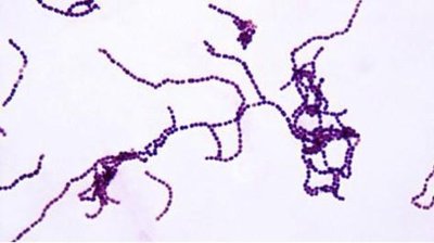
Streptococcus | Chain of cocci | ●●●● |
Staphylococcus | Cluster of cocci | ●●● ●●● |
Bacillus | Single rod | ▬ |
Streptobacillus | Chain of rods | ▬▬▬ |
Corynebacterium | Palisades | ≡≡≡ |
Complex Life Cycles of Bacteria
Phases of Bacterial Life Cycles
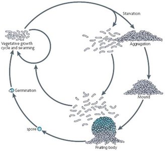
Bacteria can undergo complex life cycles, including planktonic (free-swimming), swarming, attachment, aggregation, and fruiting body formation. These adaptations allow survival in diverse environments.
Planktonic phase: Motile, free-living cells.
Attachment and aggregation: Cells adhere to surfaces and each other, forming colonies or biofilms.
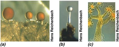
Fruiting bodies: Multicellular structures formed under starvation or stress, especially in myxobacteria.
Sporulation: Formation of resistant spores or endospores for survival under harsh conditions.
Sporulation and Endospores
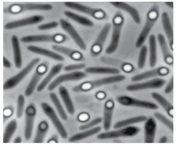
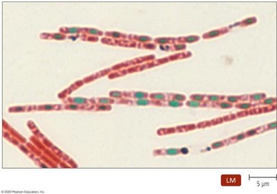
Some Gram-positive bacteria, such as Bacillus and Clostridium, form endospores that are highly resistant to environmental extremes (heat, desiccation, UV light, chemicals).
Endospores: Dormant, tough, non-reproductive structures formed within the cell.
Schaeffer-Fulton Staining: A differential stain used to visualize endospores (endospores appear green, vegetative cells red).
Myxobacteria and Actinomycetes: Complex Sporulation
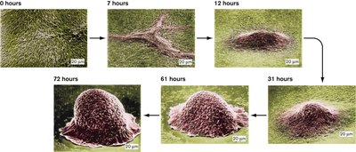
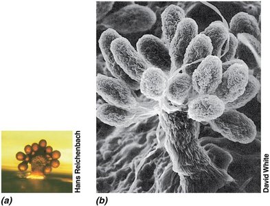
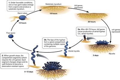
Myxobacteria (Gram-negative) and Actinomycetes (Gram-positive) exhibit complex multicellular development, including fruiting body and spore formation under starvation conditions.
Myxobacteria: Aggregate to form fruiting bodies and produce spores when nutrients are scarce.
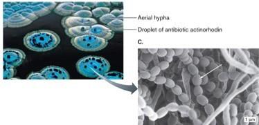
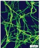
Actinomycetes: Form branching filaments and aerial mycelia, producing spores that are often mistaken for fungal spores.
Bacterial Biofilms
Biofilm Formation and Structure
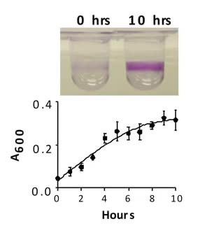
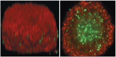
Biofilms are structured communities of bacteria adhered to surfaces and embedded in a self-produced extracellular matrix, primarily composed of polysaccharides. Biofilm development involves several stages:
Attachment: Free-floating (planktonic) cells attach to a surface.
Microcolony formation: Cells aggregate and produce extracellular polymeric substances (EPS).
Maturation: Biofilm architecture develops, with channels for nutrient flow and waste removal.
Dispersion: Cells or clusters detach to colonize new sites.
Biofilm Resistance and Medical Relevance
Bacteria in biofilms are highly resistant to antibiotics and immune responses, contributing to persistent infections, especially on medical devices (catheters, implants) and in chronic diseases (e.g., cystic fibrosis lung infections).
Medical devices: Biofilms on catheters, heart valves, and joint replacements are common sources of hospital-acquired infections.
Environmental biofilms: Legionella pneumophila in air-conditioning systems, Pseudomonas aeruginosa in water systems and wounds.
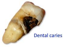
Dental plaque: A multispecies biofilm that can lead to dental caries and periodontal disease.
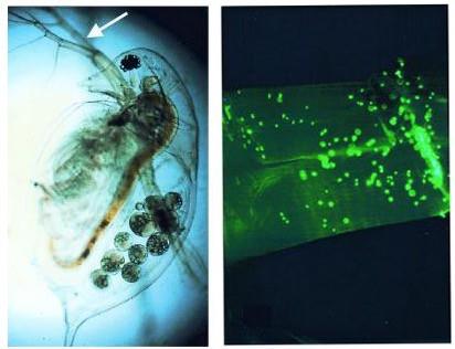
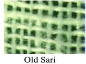
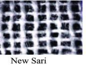
Biofilm Control: Case Study of Cholera Prevention
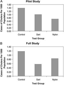
Simple filtration methods, such as using folded sari cloth, can significantly reduce waterborne diseases like cholera by trapping bacteria-laden biofilms from contaminated water.
Vibrio cholerae: Forms biofilms on “marine snow” and can be filtered out using sari cloth.
Field studies: Showed a 48% reduction in cholera cases in Bangladeshi villages using sari filtration.
Summary Table: Key Features of Prokaryotes
Feature | Bacteria | Archaea |
|---|---|---|
Cell Wall | Peptidoglycan | No peptidoglycan (varied composition) |
Membrane Lipids | Ester-linked | Ether-linked |
Genetic Material | Circular DNA, no nucleus | Circular DNA, no nucleus |
Ribosomes | 70S | 70S (distinct rRNA sequences) |
Examples | Escherichia coli, Bacillus subtilis | Halophiles, Methanogens |
Additional info: The study of prokaryotes is foundational for understanding microbial diversity, ecology, and the development of antimicrobial strategies. Their ability to form biofilms and spores is central to their survival and pathogenicity.
