 Indietro
Prokaryotic Cell Structure and Function: Microbiology Study Guide
Guida di studio - Note intelligenti
 Appunti personalizzati basati sui tuoi materiali, ampliati con definizioni chiave, esempi e contesto.
Prokaryotic Cell Basics
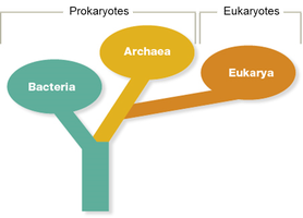
Domains of Prokaryotes
Prokaryotes are classified into two domains: Bacteria and Archaea. Both domains consist of unicellular organisms lacking a membrane-bound nucleus and other membrane-bound organelles. They are genetically simpler than eukaryotic cells. While bacteria include many human pathogens, no known human pathogens exist among archaea.
Bacteria: Found in diverse environments; many are pathogenic.
Archaea: Often inhabit extreme environments; genetically distinct from bacteria.
Similarity: Both lack membrane-bound organelles.
Difference: Archaea have unique membrane lipids and cell wall components.
Prokaryotic Cell Structure
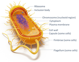
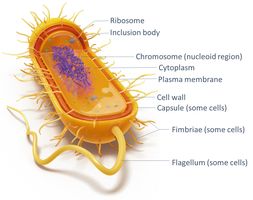
Prokaryotic cells are characterized by their simplicity and small size. Their structure includes a plasma membrane, cell wall, cytoplasm, ribosomes, nucleoid, and often external structures such as flagella and pili.
Plasma membrane: Selectively permeable boundary.
Cell wall: Provides structural support and protection.
Nucleoid: Region containing the cell's DNA.
Ribosomes: Sites of protein synthesis.
External structures: Flagella, pili, fimbriae, and glycocalyx.
Prokaryotic Cell Sizes
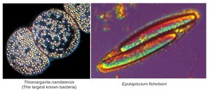
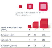
Prokaryotes are generally very small, ranging from 0.2–750 μm in diameter, with most between 0.5–2.0 μm. Their small size is due to their reliance on diffusion for nutrient uptake, which limits their surface area-to-volume ratio.
Smallest: Mycoplasma species (~300 nm).
Largest: Thiomargarita namibiensis (~750 μm).
Prokaryotic Shapes and Arrangements
Shapes
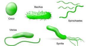
Prokaryotes exhibit a variety of shapes, which are important for identification and classification. The main shapes include:
Bacilli: Rod-shaped
Cocci: Spherical
Vibrio: Comma-shaped
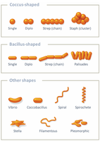
Stella: Star-shaped
Coccobacilli: Ovoid
Spirochetes: Corkscrew-shaped
Spirilla: Spiral-shaped
Arrangements
Cell division patterns result in characteristic arrangements:
Diplococci: Paired cocci
Streptococci: Chains of cocci
Staphylococci: Grapelike clusters
Diplobacilli: Paired bacilli
Streptobacilli: Chains of bacilli
Palisade: Picket fence-like clusters of bacilli
Pleomorphism
Pleomorphic bacteria can take on different forms, which may enhance their ability to cause disease by adapting to various environments.
Monomorphic: One shape
Pleomorphic: Multiple shapes
Binary Fission
Process of Binary Fission
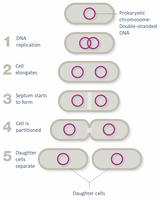
Binary fission is the primary method of reproduction in prokaryotes. It is a simple, asexual process resulting in two genetically identical daughter cells.
DNA is replicated
Cell elongates
Chromosomes are drawn to opposite ends
Septum forms at the midpoint
Septum partitions the cell, resulting in two daughter cells
Extracellular Structures
Prokaryotic Plasma Membrane
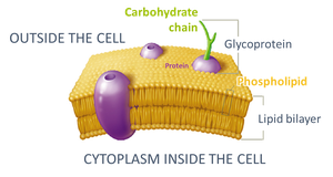
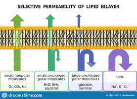
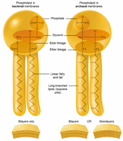
The plasma membrane is a thin, flexible phospholipid bilayer that acts as a selectively permeable barrier. In archaea, the membrane may be a monolayer, and the fatty acids are branched, unlike the linear fatty acids in bacteria.
Proteins: Function as transporters, anchors, receptors, and enzymes.
Selective permeability: Allows gases, water, and small noncharged substances to diffuse freely; ions and large polar substances require transport proteins.
Bacterial Cell Wall
The cell wall surrounds the plasma membrane and provides strength and protection. Most bacteria have a cell wall composed of peptidoglycan, a multi-layered sugar-protein complex.
Peptidoglycan: Chains of alternating sugars (NAG-NAM) cross-linked by peptides.
Exception: Mycoplasma species lack a cell wall.
Gram Staining and Cell Wall Types
Gram staining is a key clinical tool for classifying bacteria based on cell wall structure:
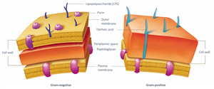
Gram-positive: Thick peptidoglycan layer, no outer membrane; stains purple.
Gram-negative: Thin peptidoglycan layer, outer membrane with lipopolysaccharide (LPS); stains pink.
Clinical Implications of Gram Status
Gram-negative bacteria are harder to kill due to their outer membrane, which acts as a selective barrier. Gram-positive bacteria are more sensitive to drugs targeting peptidoglycan and retain moisture longer. Teichoic acids in Gram-positive cell walls can promote adhesion to host tissues.
Acid-Fast Staining
Acid-fast staining detects mycolic acid in cell walls of certain genera, such as Nocardia and Mycobacterium. Acid-fast bacteria appear red/pink after staining and tend to grow slowly, requiring long, multi-drug therapies for infections.
Mycoplasma and L-Forms
Mycoplasma bacteria lack a cell wall and are pleomorphic. L-forms are bacteria that have lost their cell wall, contributing to persistent infections and resistance to environmental stresses.
Transport Mechanisms
Passive Transport
Passive transport does not require energy and includes diffusion and osmosis:
Diffusion: Movement from high to low concentration.
Simple diffusion: Small, noncharged molecules and gases.
Facilitated diffusion: Uses membrane proteins for larger or charged molecules.
Osmosis: Movement of water across a membrane from low to high solute concentration.
Effects of Osmosis
Hypertonic solution: Cell loses water; plasmolysis occurs.
Hypotonic solution: Cell gains water; osmotic lysis may occur if cell wall is damaged.
Isotonic solution: No net water movement.
Active Transport
Active transport requires energy (ATP) and can move substances against their concentration gradient. Types include:
Primary active transport: Uses ATP directly.
Secondary active transport: Uses ion gradients (symport and antiport).
Phosphotransferase system: Group translocation; transfers phosphate to transported substance.
Extracellular Structures for Movement and Adhesion
Flagella
Flagella are filament-like extensions used for motility. They rotate to propel the cell and are powered by ATP. Flagella arrangements include monotrichous (single), lophotrichous (tuft), amphitrichous (both ends), and peritrichous (all over).
Chemotaxis: Movement in response to chemicals.
Phototaxis: Movement in response to light.
Aerotaxis: Movement in response to oxygen.
Periplasmic Flagella
Found only in spirochetes, periplasmic flagella (axial filaments) are located within the periplasmic space and allow corkscrew movement through fluids.
Fimbriae and Pili
Fimbriae are short, bristle-like proteins that help bacteria adhere to surfaces. Pili are longer, more rigid, and less numerous, used for movement and DNA transfer (conjugation).
Glycocalyx
The glycocalyx is a sticky, carbohydrate-rich layer outside the cell wall. It exists as a slime layer (unorganized) or capsule (well-organized), aiding in adhesion, protection from desiccation, antibiotics, and disinfectants.
Slime layer: Loosely associated.
Capsule: Tightly associated; increases pathogenicity.
Intracellular Structures
Nucleoid
The nucleoid is the cytoplasmic region containing bacterial DNA, usually a single circular chromosome of double-stranded DNA.
Ribosomes
Prokaryotic ribosomes (70S) are nonmembranous organelles responsible for protein synthesis. Some antibiotics target the 70S ribosome, distinguishing them from eukaryotic ribosomes (80S).
Cytoskeleton
The prokaryotic cytoskeleton consists of protein filaments arranged in rings, coils, or rods, providing internal structure, support, and assisting in cell division and movement.
Inclusion Bodies
Inclusion bodies are collections of insoluble substances in the cytoplasm, often used for food storage (e.g., glycogen) or orientation (magnetic inclusion bodies).
Endospores
Endospores are metabolically inactive, dormant structures produced by certain genera (e.g., Bacillus, Clostridium, Clostridioides) to survive hostile conditions. They are not a form of reproduction but a survival mechanism. Endospores can persist for extended periods, posing challenges in healthcare settings.
Bacillus anthracis: Anthrax
Clostridium tetani: Tetanus
Clostridium botulinum: Botulism
Clostridium perfringens: Gas gangrene
Clostridioides difficile: Diarrhea and colitis
Summary Table: Gram-Positive vs. Gram-Negative Bacteria
Feature | Gram-Positive | Gram-Negative |
|---|---|---|
Peptidoglycan Layer | Thick | Thin |
Outer Membrane | Absent | Present (with LPS) |
Stain Color | Purple | Pink |
Drug Sensitivity | More sensitive to peptidoglycan-targeting drugs | More resistant due to outer membrane |
Teichoic Acids | Present | Absent |
