 Indietro
Prokaryotic Cell Structure: External and Cytoplasmic Components
Guida di studio - Note intelligenti
 Appunti personalizzati basati sui tuoi materiali, ampliati con definizioni chiave, esempi e contesto.
More External Structures of Prokaryotes
Fimbriae, Pili, and Flagella
Many bacteria and archaea possess external structures that extend beyond the cell envelope. These structures play crucial roles in protection, attachment, horizontal gene transfer, and motility.
Fimbriae: Short, thin, hairlike protein appendages (up to 1,000/cell) that mediate attachment to surfaces. Some, such as type IV fimbriae, are involved in motility or DNA uptake.
Pili: Similar to fimbriae but longer, thicker, and less numerous (1–10/cell). Sex pili are hollow and required for conjugation (gene transfer between bacteria).
Flagella: Threadlike locomotor appendages responsible for motility and swarming behavior, as well as attachment to surfaces.
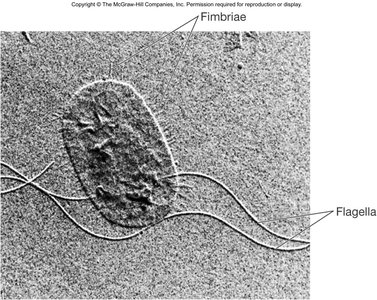
Flagella: Structure and Function
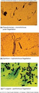
Patterns of Flagella Distribution
Flagella can be distributed in various patterns on the cell surface, which are important for bacterial identification and motility mechanisms.
Monotrichous: Single flagellum at one end.
Polar flagellum: Flagellum located at the end of the cell.
Amphitrichous: One flagellum at each end of the cell.
Lophotrichous: Cluster of flagella at one or both ends.
Peritrichous: Flagella spread over the entire surface of the cell.
Flagellar Structure
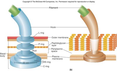
The bacterial flagellum is a complex structure composed of three main parts:
Filament: The longest part, a hollow, rigid cylinder made of flagellin protein. Some bacteria have a sheath around the filament.
Hook: A short, curved segment that links the filament to the basal body.
Basal body: A series of rings embedded in the cell envelope that function as the flagellar motor.
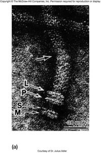
Motility in Bacteria and Archaea
Types of Motility
Prokaryotes exhibit several types of motility, allowing them to move toward favorable environments or away from harmful conditions.
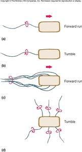
Flagellar movement: Rotation of flagella propels the cell.
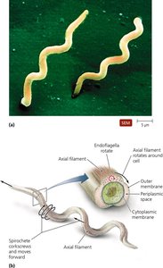
Spirochete motility: Movement via axial filaments (endoflagella) within the periplasmic space, producing a corkscrew motion.
Twitching motility: Short, jerky movements mediated by type IV pili.
Gliding motility: Smooth movement along surfaces, often involving slime secretion.
Chemotaxis and Directed Movement
Bacteria and archaea can move in response to various stimuli, a behavior known as taxis. Chemotaxis is movement toward chemical attractants (positive) or away from repellents (negative). Other forms include phototaxis (light), aerotaxis (oxygen), and magnetotaxis (magnetic fields).
Movement is regulated by chemoreceptors that detect changes in concentration of attractants or repellents.
Mechanism of Flagellar Movement
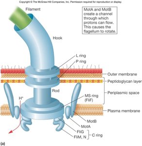
Flagellar rotation is powered by a motor embedded in the cell envelope. The motor consists of rotor and stator components:
Rotor: Composed of the C (FliG protein) ring and MS ring, which rotate and interact with the stator.
Stator: MotA and MotB proteins form channels through which protons (H+) flow, utilizing the proton motive force to generate torque and rotate the flagellum.
Spirochete Motility
Spirochetes possess endoflagella (axial filaments) located within the periplasmic space. These structures enable movement in viscous environments by producing a corkscrew motion.
Multiple flagella emanate from each end, forming an axial fibril that winds around the cell.
The corkscrew shape allows flexing and spinning movements.
Prokaryotic Cytoplasmic Structures
Cytoplasm and Cytoskeleton
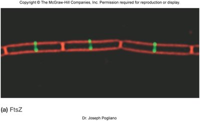
The cytoplasm is the material within the cell membrane, consisting of cytosol (90–92% water, dissolved solutes, enzymes, and ribosomes) and a cytoskeleton in some bacteria. The cytoskeleton is a network of protein fibers that plays roles in cell division, protein localization, and cell shape determination.
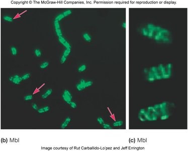
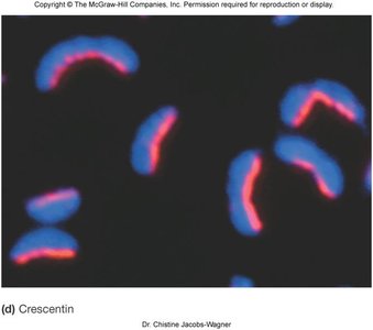
Major cytoskeletal proteins include FtsZ (tubulin homolog), MreB (actin homolog), and crescentin (intermediate filament homolog).
Intracytoplasmic Membranes
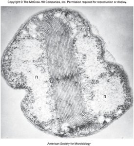
Some bacteria have internal membrane systems derived from infoldings of the plasma membrane. These structures are especially common in photosynthetic bacteria and those with high respiratory activity.
They serve as sites for ATP formation and may be analogous to thylakoids in chloroplasts.
Anammoxosomes are specialized compartments for anaerobic ammonia oxidation.
Inclusions
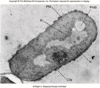
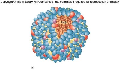
Inclusions are membrane-bound or protein-bound storage structures containing granules of organic or inorganic material. They are not infoldings but separate entities within the cytoplasm.
Functions include storage of nutrients, metabolic end products, energy, and building blocks.
Examples: Glycogen granules, poly-β-hydroxybutyrate (PHB) for carbon storage, polyphosphate (volutin) granules, and sulfur granules.
Other Inclusions
Additional types of inclusions include:
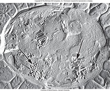
Gas vacuoles: Found in aquatic, photosynthetic bacteria and archaea, providing buoyancy via gas vesicles.
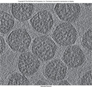
Microcompartments: Protein-based compartments for specific functions, such as carboxysomes in CO2-fixing bacteria, which contain the enzyme Rubisco for carbon fixation.
Ribosomes
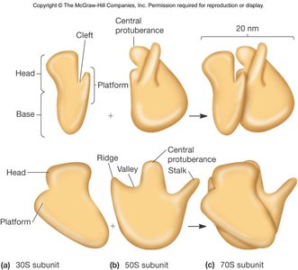
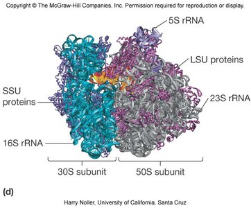
Ribosomes are complex structures composed of protein and ribosomal RNA (rRNA), serving as the sites of protein synthesis.
Bacterial and archaeal ribosomes are 70S, consisting of a 50S large subunit and a 30S small subunit.
rRNA components: 16S (small subunit), 23S and 5S (large subunit); archaea also have 5.8S rRNA.
Proteins in archaeal ribosomes are more similar to those in eukaryotes than to bacteria.
Nucleoid and Plasmids
Nucleoid
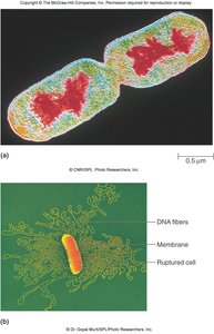
Prokaryotic cells lack a true nucleus. Instead, their DNA is located in an irregularly shaped region called the nucleoid, which typically occupies 10–15% of the cytoplasmic volume.
Most prokaryotes have a single, circular, supercoiled double-stranded DNA chromosome.
Plasmids
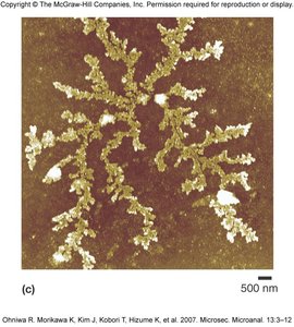
Plasmids are extrachromosomal, usually small, closed circular DNA molecules found in many bacteria, some archaea, and some fungi. They replicate independently of the chromosome and often carry non-essential genes that confer selective advantages, such as antibiotic resistance.
Episomes are plasmids that can integrate into the bacterial chromosome.
Plasmids are inherited by daughter cells but can be lost (curing).
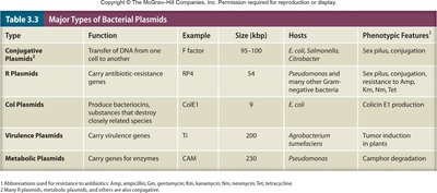
Type | Function | Example | Size (kbp) | Hosts | Phenotypic Features |
|---|---|---|---|---|---|
Conjugative Plasmids | Transfer of DNA from one cell to another | F factor | 95–100 | E. coli, Salmonella | Sex pilus, conjugation |
R Plasmids | Carry antibiotic-resistance genes | RP4 | 54 | Pseudomonas, many Gram-negative bacteria | Sex pilus, conjugation, resistance to Amp, Kan, Tetr, etc. |
Col Plasmids | Produce bacteriocins, substances that destroy closely related species | ColE1 | 6 | E. coli | Colicin E1 production |
Virulence Plasmids | Carry virulence genes | Ti | 200 | Agrobacterium tumefaciens | Tumor induction in plants |
Metabolic Plasmids | Carry genes for enzymes | CAM | 230 | Pseudomonas | Camphor degradation |
Bacterial Endospores
Endospore Formation and Structure
Some bacteria form complex, dormant structures called endospores to survive extreme environmental conditions such as heat, radiation, chemicals, and desiccation.
Sporulation: The process of endospore formation, typically initiated when growth ceases due to nutrient limitation or toxic substances. It is a complex, multistage process that can take up to 10 hours.
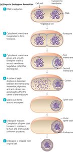
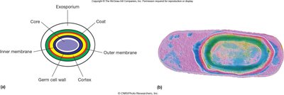
Endospore structure: The spore is surrounded by a thin exosporium, thick protein spore coat, cortex (thick peptidoglycan), and a core containing the nucleoid and ribosomes.
Germination
Germination is the process by which an endospore returns to a metabolically active, vegetative cell. This is also a complex, multistage process triggered by favorable environmental conditions.
