 Indietro
Simple Staining in Microbiology: Principles, Methods, and Applications
Guida di studio - Note intelligenti
 Appunti personalizzati basati sui tuoi materiali, ampliati con definizioni chiave, esempi e contesto.
Simple Staining
Introduction to Simple Staining
Simple staining is a fundamental microbiological technique used to enhance the visibility of bacterial cells under a microscope. By applying a single basic dye, this method allows for the observation of cell morphology, arrangement, and size, which are critical for the identification and classification of microorganisms.
Principle of Basic Stains
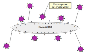
Basic stains are dyes with a positively charged chromogen (color-bearing ion).
Bacterial cells typically have a net negative charge on their surface due to the presence of acidic molecules in their cell walls.
The positively charged chromogen of the basic stain ionically bonds with the negatively charged bacterial cell, resulting in the cell taking up the color of the dye.
Purpose and Applications of Simple Staining
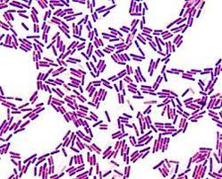
Increases contrast between the bacterial cell and the background, making cells more visible under the microscope.
Allows for the determination of cell morphology (shape), arrangement (grouping pattern), and size.
Does not differentiate between different types of bacteria (unlike differential stains such as Gram stain).
Common Basic Stains Used in Simple Staining
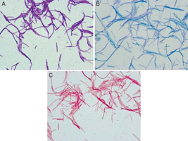
Crystal Violet
Safranin
Methylene Blue
Each of these stains can be used to color bacterial cells, but the choice of stain does not affect the basic information obtained (morphology, arrangement, size).
Bacterial Smear Preparation
Steps in Preparing a Bacterial Smear
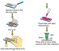
Proper smear preparation is essential for successful staining and observation. The goal is to create a thin, even layer of bacteria on the slide that will withstand the staining process.
Obtain a clean glass slide.
If using solid media, add a drop of water to the slide first.
Transfer a small amount of bacterial culture to the slide and spread it to form a thin smear.
Allow the smear to air dry completely.
Pass the slide through a flame (heat-fix) to adhere the bacteria to the slide and kill the cells.

Simple Staining Procedure
Step-by-Step Protocol
Place the prepared smear on a staining rack in a staining tray.
Flood the slide with the chosen basic stain (e.g., crystal violet, safranin, or methylene blue).
Incubate for the appropriate time (usually 30 seconds to 1 minute).
Grasp the slide with a clothespin, tilt, and rinse gently with water until the runoff is clear.
Gently blot the slide dry with bibulous paper.
Observe the stained smear under oil immersion (100x objective) using a light microscope.
Examples of Bacteria Commonly Used in Simple Staining
Bacillus subtilis
Micrococcus luteus
Staphylococcus epidermidis
These species are often used in teaching laboratories to demonstrate differences in cell morphology and arrangement.
Laboratory Application: Staining Multiple Organisms
Experimental Design
Prepare three slides, each containing smears of B. subtilis, M. luteus, and S. epidermidis.
Stain each slide with a different basic dye:
Slide 1: Crystal violet
Slide 2: Safranin
Slide 3: Methylene blue
Dispose of slides in a sharps container or keep them in a labeled blue box as instructed.
Summary Table: Simple Staining Overview
Step | Purpose | Key Points |
|---|---|---|
Smear Preparation | Adhere bacteria to slide, preserve morphology | Thin, even smear; air dry; heat fix |
Staining | Color cells for visibility | Use basic dye; incubate briefly |
Rinsing & Drying | Remove excess stain, prevent artifacts | Gentle water rinse; blot dry |
Microscopy | Observe cell morphology, arrangement, size | Oil immersion (100x objective) |
Additional info: Simple staining is a prerequisite for more advanced staining techniques, such as Gram staining and acid-fast staining, which provide further differentiation between bacterial types.
