 Indietro
Structural and Differential Stains in Microbiology: Endospore, Capsule, Flagella, and Acid-Fast Stains
Guida di studio - Note intelligenti
 Appunti personalizzati basati sui tuoi materiali, ampliati con definizioni chiave, esempi e contesto.
Structural & Differential Stains in Microbiology
Introduction
Structural and differential stains are essential techniques in microbiology for visualizing and distinguishing between different types of bacterial cells and their components. These stains help identify unique cellular structures such as endospores, capsules, flagella, and acid-fast cell walls, which are critical for classification, diagnosis, and understanding microbial physiology.
Endospore Stain
Definition and Significance
Endospores are dormant, highly resistant structures formed by genera such as Bacillus and Clostridium when environmental conditions become unfavorable.
They are resistant to heat, desiccation, chemicals, and UV light, allowing survival for extended periods.
Endospores germinate into vegetative cells when conditions improve.
Schaeffer-Fulton Endospore Staining Method
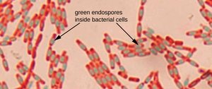
Primary stain: Malachite green (driven into endospores by steam heating)
Decolorizer: Water (removes stain from vegetative cells, not endospores)
Counterstain: Safranin (stains vegetative cells pink/red)
Results: Endospores appear green; vegetative cells appear pink/red.
Example organisms: Bacillus subtilis, Bacillus megaterium (positive); Escherichia coli, Enterobacter aerogenes (negative)
Procedure Summary
Prepare and heat-fix bacterial smears.
Cover with malachite green and steam for 5 minutes.
Rinse with water to decolorize vegetative cells.
Counterstain with safranin for 30 seconds.
Rinse, dry, and observe under oil immersion.
Capsule Stain
Definition and Significance
Capsules are organized layers of polysaccharide or protein surrounding some bacteria, firmly attached to the cell wall.
If loosely attached, the structure is called a slime layer.
Capsules prevent phagocytosis, serving as important virulence factors.
Capsule Staining Technique
Negative stain: Capsule does not take up stain.
Primary stain: Congo red (acidic dye) stains the background red.
Counterstain: Acid fuchsin stains the bacterial cells red.
Result: Capsule appears as a clear halo around the red cell against a red background.
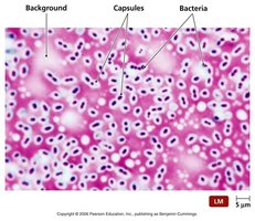
Example organisms: Streptococcus salivarius (capsule-positive), Enterobacter aerogenes (capsule-negative or minimal)
Flagella Stain
Definition and Significance
Flagella are thin, proteinaceous appendages that provide motility to bacteria.
They originate in the cytoplasm and project through the cell wall.
Flagella are too thin to be seen with standard light microscopy; a mordant is used to coat and thicken them for visualization.
Motility aids in colonization and infection of host tissues.
Flagellar Arrangements
Monotrichous: One flagellum at one end
Lophotrichous: Multiple flagella at one end
Amphitrichous: Flagella at both ends
Peritrichous: Flagella distributed all around the cell
Example organisms: Proteus vulgaris, Enterobacter aerogenes, Bacillus megaterium, Escherichia coli (all peritrichous)
Acid-Fast Stain
Definition and Significance
Acid-fast bacteria have cell walls rich in mycolic acid, a waxy lipid that makes the wall nearly impermeable to most stains.
This property is characteristic of Mycobacterium (e.g., tuberculosis, leprosy) and some Nocardia species.
Acid-Fast Staining Methods
Ziehl-Neelsen method: Uses heat to drive carbol fuchsin into the cell wall.
Kinyoun modification: Cold stain with higher phenol concentration; no heat required (safer for teaching labs).
Staining Results
Primary stain: Carbol fuchsin (stains acid-fast cells pink/red)
Decolorizer: Acid-alcohol (removes stain from non-acid-fast cells)
Counterstain: Methylene blue (stains non-acid-fast cells blue)
Result: Acid-fast cells = pink/red; non-acid-fast cells = blue
Example organisms: Mycobacterium smegmatis (acid-fast positive), Escherichia coli (acid-fast negative)
Summary Table: Key Structural and Differential Stains
Stain | Primary Stain | Counterstain | Positive Result | Negative Result | Key Structure Detected |
|---|---|---|---|---|---|
Endospore | Malachite green | Safranin | Green endospores | Red vegetative cells only | Endospores |
Capsule | Congo red | Acid fuchsin | Clear halo (capsule) | No halo | Capsule |
Acid-Fast | Carbol fuchsin | Methylene blue | Pink/red cells | Blue cells | Mycolic acid-rich cell wall |
Key Terms and Concepts
Primary stain: The first dye applied in a differential staining procedure, targeting the structure of interest.
Counterstain: A secondary dye that stains cells or structures not retained by the primary stain, providing contrast.
Decolorizer: A reagent that removes the primary stain from certain cell types or structures.
Mordant: A substance that increases the affinity of a stain for a structure (e.g., used in flagella staining).
Practice Questions
What are the primary and counterstains used in acid-fast staining?
What color indicates a positive acid-fast test, and what macromolecule is responsible?
What are the primary and counterstains used in endospore staining?
What do the red structures represent in endospore staining?
What two stains are used in capsule staining?
List and define the four flagellar arrangements.
Additional info: The above notes integrate both procedural and conceptual knowledge, providing a comprehensive overview suitable for exam preparation in a college-level microbiology course.
