 Indietro
The Prokaryote Cell Envelope and Growth: Structure, Function, and Clinical Relevance
Guida di studio - Note intelligenti
 Appunti personalizzati basati sui tuoi materiali, ampliati con definizioni chiave, esempi e contesto.
The Prokaryote Cell Envelope
Overview of the Prokaryote Envelope
The cell envelope is a complex, multi-layered structure that surrounds the cytoplasm of prokaryotic cells, providing structural support, protection, and mediating interactions with the environment. The composition and organization of the envelope vary significantly among different groups of prokaryotes, including Bacteria and Archaea.
Monoderm bacteria: Possess a single cell membrane, typically Gram-positive.
Diderm bacteria: Possess two membranes (inner and outer), typically Gram-negative.
Archaea: Exhibit unique envelope structures distinct from Bacteria.
Gram-Positive vs. Gram-Negative Cell Envelopes
Gram-positive and Gram-negative bacteria differ fundamentally in their cell envelope architecture, which influences their staining properties, susceptibility to antibiotics, and ecological roles.
Gram-positive bacteria have a thick, multilayered peptidoglycan cell wall and lack an outer membrane.
Gram-negative bacteria have a thin peptidoglycan layer and an additional outer membrane containing lipopolysaccharides (LPS).
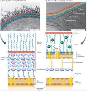
S-layer: A proteinaceous surface layer found in many Gram-positive and Gram-negative bacteria, as well as Archaea. Functions include protection, surface adhesion, and acting as a molecular sieve.
The Cell Membrane(s)
Structure and Function of the Cell Membrane
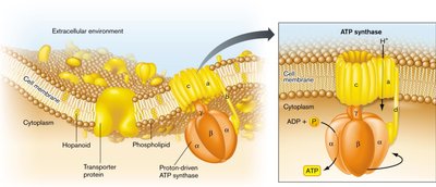
The cell membrane is a phospholipid bilayer embedded with proteins, serving as a selective barrier and site for energy generation and transport processes.
Phospholipids form the basic structure of the membrane, with hydrophilic heads and hydrophobic tails.
Membrane proteins facilitate ion and metabolite transport, signal detection, catalysis, and structural support.
ATP synthase utilizes proton gradients to synthesize ATP, the universal energy currency of the cell.
Phospholipid Diversity
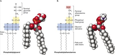
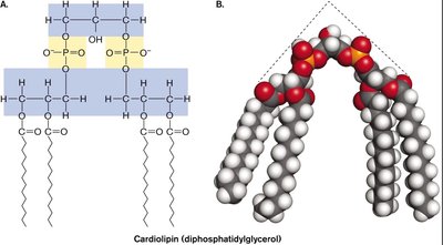
Phospholipids vary in their head groups and fatty acid composition, influencing membrane fluidity and function.
Phosphatidylglycerol and phosphatidylethanolamine are common bacterial phospholipids.
Cardiolipin (diphosphatidylglycerol) is enriched at cell poles and division sites.
Archaeal Membrane Lipids
Archaeal membranes are chemically distinct from bacterial and eukaryotic membranes, contributing to their ability to thrive in extreme environments.
Archaeal lipids are based on L-glycerol diether or tetraether backbones with isoprenoid side chains and ether linkages.
Bacterial and eukaryotic membranes use D-glycerol with fatty acid side chains and ester linkages.
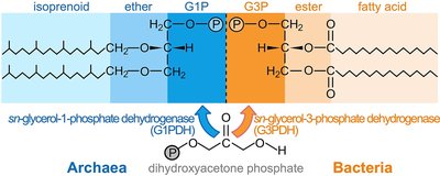
The Bacterial Cell Wall
Peptidoglycan Structure and Synthesis
The bacterial cell wall is primarily composed of peptidoglycan (murein), a polymer of sugars and amino acids that provides rigidity and shape to the cell.
Peptidoglycan is unique to bacteria and is a major target for antibiotics.
Synthesis involves penicillin-binding proteins and cytoskeletal elements, which cross-link the sugar chains with peptides.
Classic antibiotics (e.g., penicillin) inhibit these proteins, preventing cell wall formation and leading to cell lysis.

Variations in Cell Envelope Structure
Gram-positive and Gram-negative bacteria differ in the thickness and organization of their peptidoglycan layer and the presence of outer membrane components.
Gram-positive: Thick peptidoglycan, teichoic acids, no outer membrane.
Gram-negative: Thin peptidoglycan, outer membrane with LPS, periplasmic space.
Clinical Relevance: Gram Staining and Antibiotic Selection
Gram staining is a critical diagnostic tool that distinguishes bacteria based on cell wall structure, guiding empirical antibiotic therapy.
Gram-positive bacteria stain purple due to thick peptidoglycan retaining crystal violet dye.
Gram-negative bacteria stain pink/red due to thin peptidoglycan and outer membrane allowing dye to wash out.
Some bacteria (e.g., Mycobacterium) are Gram-neutral due to unique envelope composition.
Bacterial Motility: The Flagellum
Structure and Function of the Bacterial Flagellum
The bacterial flagellum is a rotary motor apparatus that enables motility in liquid environments. It is anchored in the cell envelope and powered by a proton (H+) gradient.
Composed of a basal body, hook, and filament.
Rotation can be clockwise (CW) or counterclockwise (CCW), allowing directional movement.
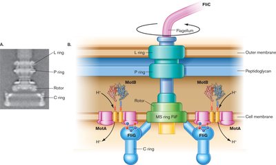
Flagellar Arrangements
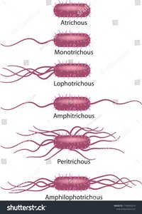
Bacteria exhibit various flagellar arrangements, which influence their motility patterns.
Monotrichous: Single flagellum at one pole.
Lophotrichous: Tuft of flagella at one pole.
Amphitrichous: Single flagellum at both poles.
Peritrichous: Flagella distributed over the entire cell surface.
Amphilophotrichous: Tuft of flagella at both poles.
Bacterial vs. Archaeal Flagella
Although both Bacteria and Archaea possess motility structures, their flagella (archaellum in Archaea) differ in origin, structure, and energy source.
Bacterial flagellum: Hollow filament, powered by proton or sodium gradient, assembled at the tip.
Archaeal archaellum: Solid filament, powered by ATP hydrolysis, assembled at the base.
Cell Growth and Division
Mechanisms of Cell Division
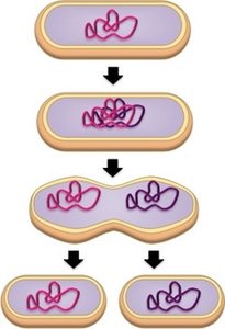
Prokaryotic cell division is primarily achieved through binary fission, resulting in two genetically identical daughter cells. Some bacteria divide by budding.
FtsZ: Forms a ring at the cell center, guiding septum formation.
MreB: Guides cell elongation by organizing peptidoglycan synthesis.
Crescentin: Promotes curved cell shapes in certain bacteria.
Growth Curve and Phases
Bacterial populations in batch culture exhibit a characteristic growth curve with four main phases:
Lag Phase: Cells adapt to new environment; no population growth.
Log (Exponential) Phase: Rapid cell division; generation time is measured here.
Stationary Phase: Growth ceases due to nutrient depletion and waste accumulation; stress response genes are upregulated.
Death Phase: Viable cell numbers decline, often exponentially.
Environmental Factors Affecting Growth
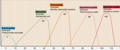
Abiotic factors such as temperature, pH, osmolarity, oxygen/CO2 levels, and light influence microbial growth rates and survival.
Optimal growth temperatures vary widely among prokaryotes, from psychrophiles (<4°C) to hyperthermophiles (>100°C).
Membrane Transport and Nutrient Uptake
Types of Membrane Transport
Prokaryotes utilize various mechanisms to transport nutrients and ions across their membranes:
Passive transport: Movement down a concentration gradient without energy input.
Active transport: Requires energy (ATP or ion gradients) to move substances against their gradient.
ABC (ATP-Binding Cassette) transporters: Use ATP hydrolysis to import nutrients.
Symport and antiport: Coupled transport mechanisms using ion gradients.
Macronutrients and Micronutrients
Microbial cells require macronutrients (C, H, O, N, S, P, K+, Mg2+, Ca2+) in large amounts and micronutrients (Zn2+, Mn2+, Mo2+) in trace amounts for growth and metabolism.
Summary Table: Gram-Positive vs. Gram-Negative Bacteria
Bacteria | Commonly found in | Common treatment |
|---|---|---|
Gram-Positive | ||
Staphylococci, Streptococci | Skin and wound infections | Vancomycin, Teicoplanin, Gentamicin (Staphylococci); Resistant to: Cephalosporins, metronidazole (except clostridia) |
Staph, Enterococcus, corynebacteria | Urine related infection | |
Clostridia | Gangrenous wound infection, abdominal infections | |
Gram-Negative | ||
The Coliform bacteria (E. coli, Enterobacter, Salmonella) | The GUT UTI, some wound infection, septicaemia | Gentamicin, ciprofloxacin, cephalosporins, amoxicillin, trimethoprim; Resistant to: Amoxicillin, most other AB's |
Pseudomonas | Most common: chronic leg ulcers, pneumonia, chest infection after flu | |
Bacteroides – anaerobic bacteria | Abdominal infection, soft tissue infection below the waist | Metronidazole, clindamycin; Resistant to: Benzylpenicillin, amoxicillin, cephalosporins, gentamicin, macrolides |
Additional info: Table summarizes common pathogens, infection sites, and antibiotic resistance patterns for Gram-positive and Gram-negative bacteria.
