 Indietro
Chapter 7:Viruses and Prions: Structure, Classification, and Multiplication- Book Notes
Guida di studio - Note intelligenti
 Appunti personalizzati basati sui tuoi materiali, ampliati con definizioni chiave, esempi e contesto.
Viruses and Prions
Introduction to Viruses
Viruses are extremely abundant and infect every type of cell, including bacteria, algae, fungi, protozoa, plants, and animals. They are best described as active or inactive rather than alive or dead, since they cannot multiply independently outside a host cell. Viruses are obligate intracellular parasites, requiring a host cell's genetic and metabolic machinery to replicate.
Not living: Most viruses cannot multiply independently from the host cell.
Influence on evolution: Viruses have shaped the genetic makeup of their hosts, and a significant portion of the human genome consists of remnants of ancient viral infections.
Part of microbiome: Viruses are a normal part of our microbiome.
Properties of Viruses
Not cells: Viruses lack cellular structure.
Obligate intracellular parasites: Infect bacteria, protozoa, fungi, algae, plants, and animals.
Inactive outside host: Only active inside host cells.
Basic structure: Protein shell (capsid) surrounding nucleic acid core.
Ultramicroscopic: Size ranges from 20 nm to 1,500 nm.
Genetic material: DNA or RNA, but never both; can be double- or single-stranded.
Surface molecules: Determine specificity for attachment to host cell.
Replication: Control host cell's genetic material to synthesize and assemble new viruses.
Lack metabolic enzymes: Usually lack enzymes for most metabolic processes and machinery for making proteins.
Viral Size Range
Viruses are much smaller than bacteria and other microorganisms. Electron microscopes are required to detect them.
Parvoviruses: 0.02 μm in diameter
Pandoravirus: As large as Streptococcus bacterium
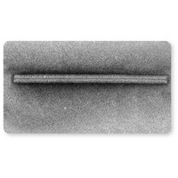
Cylindrical viruses: 0.8 μm long, 0.015 μm in diameter

Methods of Viewing Viruses
Viruses are visualized using electron microscopy and special stains, as they are too small for light microscopy.
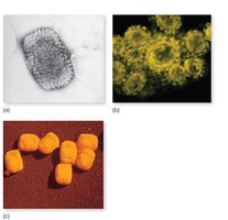
General Structure of Viruses
Virus Particle Structure
The simplest virus structure consists of a piece of genetic material (DNA or RNA) in the central core, covered by a protein coat (capsid). Some viruses also contain one or two enzymes.
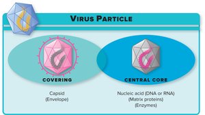
Viral Components
Capsid: Protein shell surrounding the nucleic acid.
Nucleocapsid: Capsid and nucleic acid together.
Envelope: Modified piece of host cell membrane; not present in all viruses.
Naked viruses: Viruses lacking an envelope.
Spikes: Surface proteins projecting from the nucleocapsid or envelope, allowing attachment to host cells.
Generalized Structure of Viruses
A virion is a fully formed virus able to establish infection in a host.
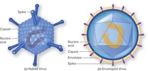
The Viral Capsid: Protective Outer Shell
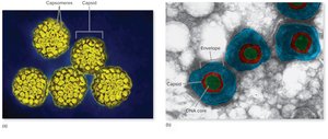
Capsomeres: Identical protein subunits that self-assemble to form the capsid.
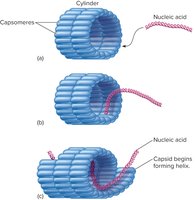
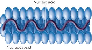
Helical capsid: Rod-shaped capsomeres form a continuous helix around the nucleic acid.
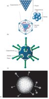
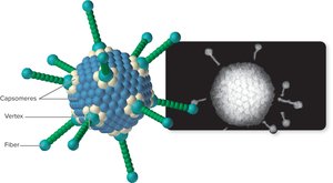
Icosahedral capsid: Three-dimensional, 20-sided figure with 12 evenly spaced corners.
Helical Nucleocapsids
Helical capsids are formed by rod-shaped capsomeres bonding together to create hollow discs, which link to form a continuous helix. The nucleic acid is coiled within this helix.
Icosahedral Capsids
Icosahedral capsids are 20-sided structures with 12 corners. The arrangement and number of capsomeres vary among viruses.
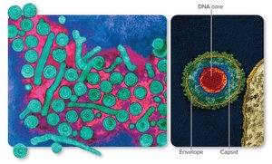
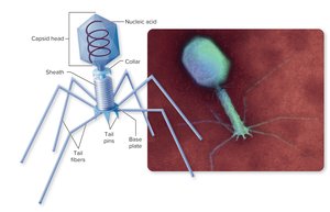
Complex Capsids
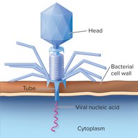
Complex capsids are found in bacteriophages and are composed of multiple types of proteins, taking shapes that are not symmetrical. They are never enveloped.
Nonenveloped and Enveloped Viruses
Viruses can be classified as nonenveloped (naked) or enveloped, depending on the presence of a lipid membrane derived from the host cell.
The Viral Envelope
Composition: Derived from the host cell membrane or nuclear membrane.
Viral proteins: Replace regular membrane proteins.
Spikes: Glycoproteins essential for attachment to the host cell.
Nucleic Acids: At the Core of a Virus
The viral genome consists of either DNA or RNA, but not both. The number of viral genes is usually small compared to cellular organisms, and viruses possess only the genes necessary to invade host cells and redirect their activity.
Positive-sense RNA: Single-stranded RNA genomes ready for immediate translation into proteins.
Negative-sense RNA: RNA genomes that must be converted into the proper form to be made into proteins.
Viral Classification and Naming
Classification of Viruses
Informal classification: Based on host (animal, plant, bacterial), envelope presence, nucleic acid type, and capsid shape.
Formal classification: Based on structure, chemical composition, and genetic makeup.
Orders: Suffix –virales
Families: Suffix –viridae
Genera: Suffix –virus
Multiplication Cycle of Animal Viruses
Six-Step Multiplication Cycle
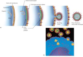
The multiplication cycle of animal viruses consists of six general phases: adsorption, penetration, uncoating, synthesis, assembly, and release.
Adsorption: Virus attaches to specific receptor sites on the host cell membrane.
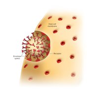
Penetration and Uncoating: Virus enters the cell via endocytosis or fusion, and the envelope and capsid are dissolved to release the viral genome.
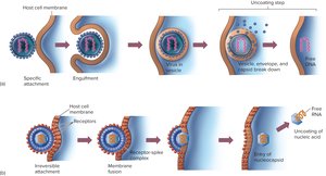
Synthesis: Viral nucleic acid takes control of the host's synthetic and metabolic machinery. RNA viruses replicate in the cytoplasm; DNA viruses replicate in the nucleus.
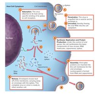
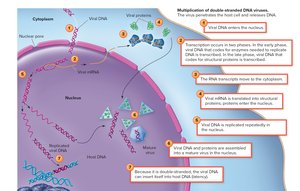
Assembly: New viral components are assembled into mature virions.
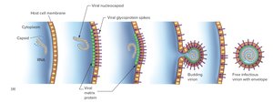
Release: Enveloped viruses are released by budding or exocytosis, while nonenveloped viruses are released by cell lysis.
Damage to the Host Cell
Cytopathic Effects (CPEs): Virus-induced damage alters cell appearance, causing disorientation, changes in shape or size, and intracellular damage.
Inclusion bodies: Compacted masses of viruses or damaged cell organelles.
Syncytia: Fusion of multiple host cells into single large cells with multiple nuclei.
Persistent and Transforming Infections
Persistent infections: Cell harbors the virus without immediate lysis; viral DNA may be incorporated into host DNA (provirus).
Chronic latent state: Periodic reactivation after a period of inactivity.
Oncogenic viruses: Viruses that cause cancer by carrying genes or producing proteins that induce loss of growth regulation.

Bacteriophages: Viruses That Infect Bacteria
Bacteriophage Structure and Life Cycle
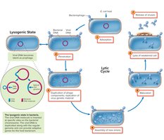
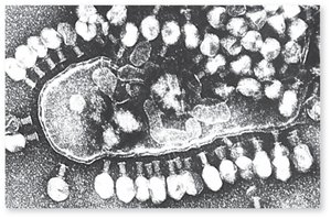
Bacteriophage: Viruses that infect bacteria; most contain dsDNA.
Lytic cycle: Ends in destruction of the bacterial cell.
Lysogenic cycle: Phage DNA is incorporated into the host cell DNA as a prophage.
Lysogenic conversion: Acquisition of new traits from temperate phage, sometimes resulting in toxin production.
Techniques in Cultivating and Identifying Animal Viruses
Viral Cultivation Methods
In vivo: Cultivation in lab animals or embryonic bird tissues.
In vitro: Cultivation in cell or tissue culture.
Primary purposes: Isolate and identify viruses, prepare viruses for vaccines, research viral structure and effects.
Using Bird Embryos
Bird embryos provide a sterile environment and multiple tissues for viral multiplication.

Viruses and Human Health
Importance and Challenges
Acute infections: Most common cause of acute infections (colds, chickenpox, influenza, herpes, warts).
High mortality: Some viruses (rabies, Ebola) have high mortality rates.
Long-term debility: Polio, neonatal rubella.
Therapies: Rapid mutation rates make antiviral drug design difficult; vaccines are preferred.
Interferon: Naturally occurring human cell product used to prevent and treat viral infections.
Prions and Other Noncellular Infectious Agents
Prions
Spongiform encephalopathies: Chronic, persistent diseases in humans and animals; brain tissue resembles a sponge.
Prions: Infectious protein fibrils deposited in brain tissue; lack nucleic acid.
Diseases: Creutzfeldt-Jakob Disease (CJD) in humans, scrapie in sheep, bovine spongiform encephalopathy in cows.
Satellite Viruses and Viroids
Satellite viruses: Dependent on other viruses for replication (e.g., adeno-associated virus).
Viroids: Virus-like agents that parasitize plants; composed only of naked RNA strands.
Example: Viroids are significant pathogens in tomatoes, potatoes, cucumbers, citrus trees, and chrysanthemums.
