 Indietro
Viruses, Viroids, and Prions: Structure, Classification, and Replication
Guida di studio - Note intelligenti
 Appunti personalizzati basati sui tuoi materiali, ampliati con definizioni chiave, esempi e contesto.
Characteristics of Viruses
Definition and General Properties
Viruses are minuscule, acellular infectious agents that contain either DNA or RNA as their genetic material. They are responsible for a wide range of diseases in humans, animals, plants, and even bacteria. Viruses are unique in that they are not considered living organisms by all scientists, as they lack cellular structure and independent metabolism.
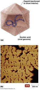
Extracellular State (Virion): The virus exists as a complete particle, with a protein coat (capsid) surrounding its nucleic acid. Some virions possess a phospholipid envelope derived from the host cell membrane.
Intracellular State: Once inside a host cell, the capsid is removed, and the virus exists as naked nucleic acid.
Are Viruses Alive?
The debate over whether viruses are alive centers on their lack of cellular structure and metabolism. Some scientists view them as complex pathogenic chemicals, while others consider them the simplest forms of life due to their ability to invade cells, control host machinery, and replicate.
Genetic Material of Viruses
Nature and Diversity of Viral Genomes
Viruses exhibit greater diversity in their genetic material than cellular organisms. Their genomes may be composed of DNA or RNA, but never both. This diversity is a primary criterion for virus classification.
Types of Viral Genomes: Double-stranded DNA (dsDNA), single-stranded DNA (ssDNA), double-stranded RNA (dsRNA), or single-stranded RNA (ssRNA).
Classification: The type of nucleic acid, presence of an envelope, shape, and size are used to classify viruses.
Hosts of Viruses
Host Specificity
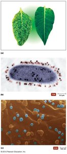
Most viruses are highly specific, infecting only particular cells of a specific host species. Some, known as generalists, can infect multiple cell types or species.
Size and Morphology of Viruses
Relative Size of Viruses
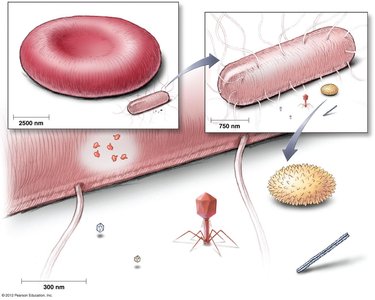
Viruses are much smaller than most cells and organelles. Their size typically ranges from 20 nm to 300 nm, making them visible only with electron microscopy.
Capsid Morphology and Viral Shapes
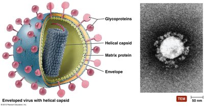
The capsid is a protein shell that protects the viral genome and facilitates attachment to host cells. Viruses exhibit three basic shapes:
Helical: Rod-shaped, with the genome coiled inside a helical capsid.
Polyhedral: Many-sided, often icosahedral.
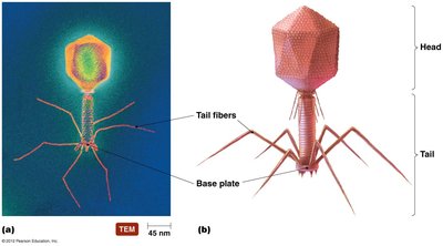
Complex: Structures that do not fit into the other categories, such as bacteriophages.

The Viral Envelope
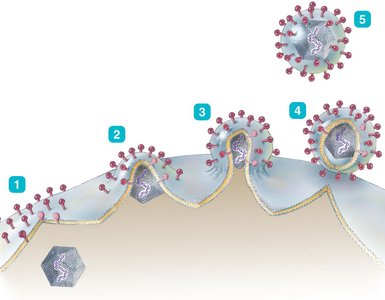
Some viruses possess an envelope derived from the host cell membrane during replication or release. This envelope contains viral proteins and glycoproteins that play a crucial role in host recognition and attachment.
Classification of Viruses
Taxonomic Criteria
Viruses are classified based on:
Type of nucleic acid (DNA or RNA, single or double-stranded)
Presence or absence of an envelope
Shape (helical, polyhedral, complex)
Size
Viral genera are grouped into families, but relationships among viruses are not as well understood as in cellular organisms.
Family | Strand Type | Representative Genera (Diseases) |
|---|---|---|
Poxviridae | Double | Orthopoxvirus (smallpox) |
Herpesviridae | Double | Simplexvirus (herpes), Varicellovirus (chickenpox), Epstein-Barr virus (mononucleosis, Burkitt’s lymphoma), Cytomegalovirus (birth defects), Roseolovirus (roseola) |
Papillomaviridae | Double | Papillomavirus (warts, cervical and penile cancers) |
Polyomaviridae | Double | Polyomavirus (progressive multifocal leukoencephalopathy) |
Adenoviridae | Double | Mastadenovirus (conjunctivitis, respiratory infections) |
Hepadnaviridae | Partial single and partial double | Orthohepadnavirus (hepatitis B) |
Parvoviridae | Single | Erythrovirus (erythema infectiosum) |
Viral Replication
Entry into Host Cells
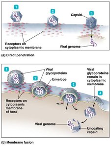
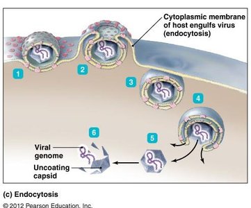
Viruses enter host cells through several mechanisms:
Direct Penetration: The viral genome is injected directly into the host cell (e.g., poliovirus).
Membrane Fusion: The viral envelope fuses with the host membrane, releasing the capsid (e.g., HIV, measles, SARS-CoV-2).
Endocytosis: The host cell engulfs the virus in a vesicle (e.g., adenovirus, herpesvirus).
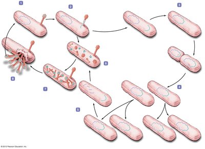
Lytic Replication Cycle
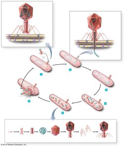
The lytic cycle is a replication process that results in the destruction (lysis) of the host cell. It consists of five stages:
Attachment
Entry
Synthesis
Assembly
Release
Lysogenic Replication Cycle
Some bacteriophages undergo lysogeny, where the viral genome integrates into the host chromosome and replicates along with it. The host cell survives and divides, passing the viral DNA to daughter cells. Environmental triggers can induce the lytic cycle.
Replication of Animal Viruses
Animal viruses follow similar replication steps as bacteriophages but differ due to the presence of envelopes, the eukaryotic nature of host cells, and the absence of a cell wall. Attachment is mediated by glycoprotein spikes or other molecules. DNA viruses often replicate in the nucleus, while RNA viruses replicate in the cytoplasm.
Latency in Animal Viruses
Some animal viruses can remain dormant (latent) within host cells for years. Latent viruses may or may not integrate into the host genome. If integrated, the provirus state is permanent.
The Role of Viruses in Cancer
Oncogenic Viruses
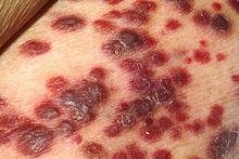
Some viruses can induce cancer by disrupting normal cell cycle regulation. They may insert oncogenes or disrupt tumor suppressor genes, leading to uncontrolled cell division (neoplasia). Viruses are implicated in 20–25% of human cancers, including Burkitt’s lymphoma, Hodgkin’s disease, Kaposi’s sarcoma, cervical cancer, and liver cancer.
Culturing Viruses in the Laboratory
Methods of Culturing Viruses
In Mature Organisms: Viruses can be grown in bacteria, plants, or animals.
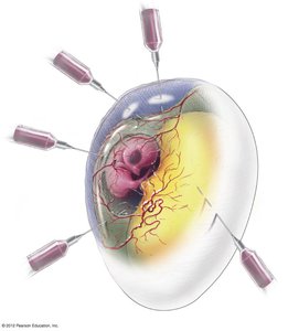
In Embryonated Chicken Eggs: Eggs provide a sterile, nutrient-rich environment for viral growth and are commonly used for vaccine production.
In Cell (Tissue) Culture: Cells isolated from organisms are grown in vitro. Two types of cultures are used: diploid cell cultures (from normal cells) and continuous cell cultures (from tumor cells).

Other Parasitic Particles: Viroids and Prions
Viroids
Viroids are extremely small, circular pieces of RNA that are infectious and pathogenic in plants. Unlike viruses, viroids lack a protein capsid.
Prions
Prions are infectious proteins that cause fatal neurodegenerative diseases. They induce abnormal folding of normal cellular proteins, leading to brain damage characterized by spongiform changes. Prion diseases are resistant to standard decontamination methods and can only be destroyed by incineration or autoclaving in 1 M NaOH.
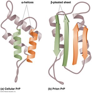
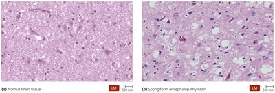
Additional info: Prion diseases include Creutzfeldt-Jakob disease, mad cow disease (bovine spongiform encephalopathy), and scrapie in sheep.
