 Indietro
Viruses, Viroids, and Prions: Structure, Classification, and Replication
Guida di studio - Note intelligenti
 Appunti personalizzati basati sui tuoi materiali, ampliati con definizioni chiave, esempi e contesto.
Viruses, Viroids, and Prions
General Characteristics of Viruses
Viruses are unique infectious agents that differ fundamentally from bacteria and other microorganisms. They are obligate intracellular parasites, meaning they require living host cells to multiply. Viruses contain a single type of nucleic acid (either DNA or RNA), a protein coat called a capsid, and sometimes an envelope derived from the host cell membrane. They lack ribosomes and ATP-generating mechanisms, making them entirely dependent on host cellular machinery for replication.
Obligate intracellular parasites: Cannot reproduce outside a host cell.
Genome: Contains either DNA or RNA, never both.
Capsid: Protein coat that protects the nucleic acid.
Envelope: Lipid, protein, and carbohydrate layer present in some viruses.
No ribosomes or ATP-generating mechanisms: Cannot synthesize proteins or generate energy independently.
Viruses and Bacteria Compared
Viruses differ from bacteria in several key aspects, including their structure, replication, and sensitivity to antibiotics and interferons. The following table summarizes these differences:
Feature | Typical Bacteria | Rickettsias/Chlamydias | Viruses |
|---|---|---|---|
Intracellular Parasite | No | Yes | Yes |
Plasma Membrane | Yes | Yes | No |
Binary Fission | Yes | Yes | No |
Pass through Bacteriological Filters | No | No/Yes | Yes |
Possess Both DNA and RNA | Yes | Yes | No |
ATP-Generating Metabolism | Yes | Yes/No | No |
Ribosomes | Yes | Yes | No |
Sensitive to Antibiotics | Yes | Yes | No |
Sensitive to Interferon | No | No | Yes |
Host Range and Viral Size
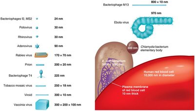
The host range of a virus is the spectrum of host cells it can infect, determined by specific attachment sites and cellular factors. Most viruses infect only specific cell types within a single host species. Viruses are extremely small, ranging from 20 nm to 1000 nm in length, which allowed their discovery before the invention of the electron microscope due to their ability to pass through filters that retain bacteria.
Viral Structure and Morphology
Basic Viral Structure
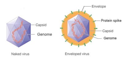
A complete, fully developed viral particle is called a virion. The main components of a virion include:
Nucleic acid: DNA or RNA, single- or double-stranded, linear or circular.
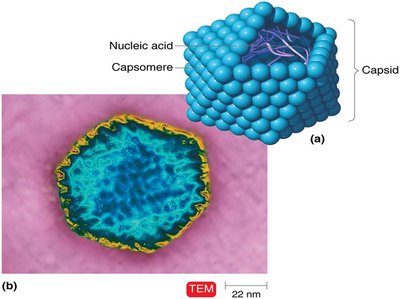
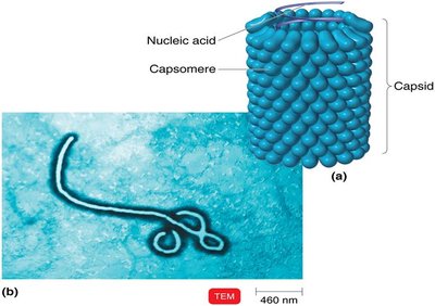
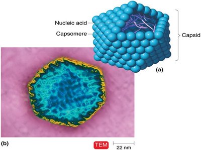
Capsid: Protein coat made of subunits called capsomeres.
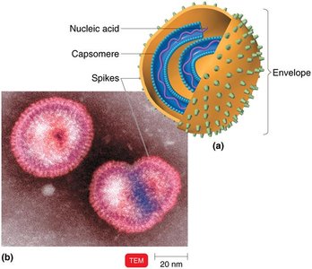
Envelope: Lipid, protein, and carbohydrate coating present in some viruses.
Spikes: Glycoprotein projections from the outer surface, important for attachment to host cells.
Viral Morphology
Viruses exhibit several morphological types:
Helical viruses: Hollow, cylindrical capsid (e.g., Ebola virus).
Polyhedral viruses: Many-sided, typically icosahedral (e.g., adenovirus).
Enveloped viruses: Surrounded by a lipid envelope (e.g., influenza virus).
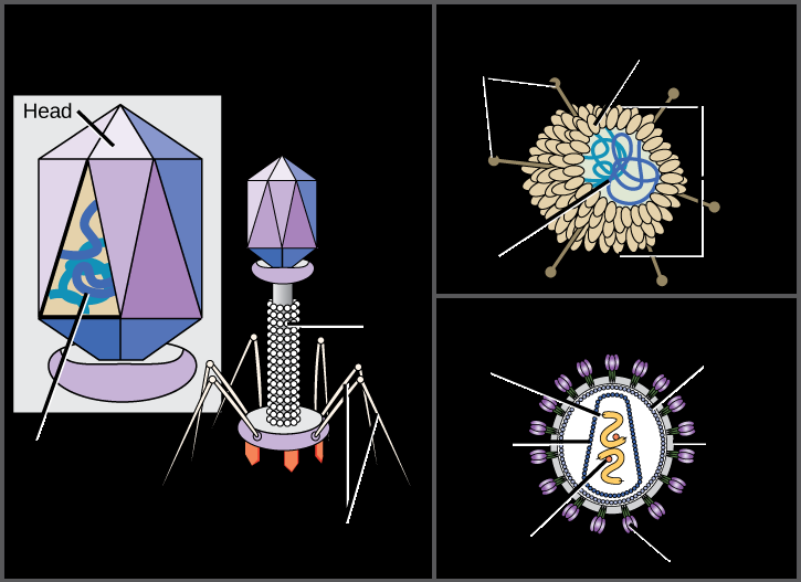
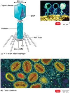
Complex viruses: Complicated structures, such as bacteriophages with a head, tail, and tail fibers.
Taxonomy of Viruses
Classification and Nomenclature
Viruses are classified based on their genetic material, structure, and host range. The taxonomy uses the following conventions:
Genus names: End in -virus
Family names: End in -viridae
Order names: End in -ales
Viral species: Group of viruses sharing the same genetic information and ecological niche (host)
Subspecies: Designated by a number
Isolation, Cultivation, and Identification of Viruses
Growing Bacteriophages
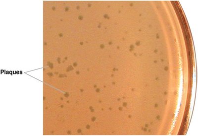
Bacteriophages are grown in bacteria, forming plaques (clearings) on a lawn of bacteria on agar plates. Each plaque corresponds to a single virus and can be quantified as plaque-forming units (PFU).
Growing Animal Viruses
In living animals: Used to study immune responses.
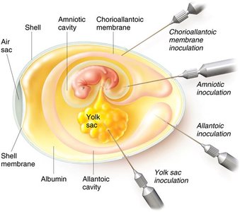
In embryonated eggs: Virus is injected into various sites; growth is indicated by embryo changes or death.
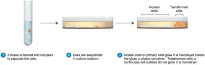
In cell cultures: Tissues are treated with enzymes to separate cells, which are then grown in culture. Virally infected cells show cytopathic effects (CPE).
Viral Identification
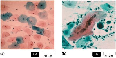
Cytopathic effects: Observable changes in cell morphology due to viral infection.
Serological tests: Detection of viral proteins using antibodies (e.g., Western blotting).
Nucleic acid techniques: RFLP (Restriction Fragment Length Polymorphism), PCR (Polymerase Chain Reaction).
Viral Multiplication
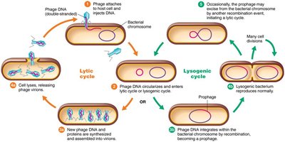
Lytic and Lysogenic Cycles in Bacteriophages
Bacteriophages can replicate via two main cycles:
Lytic cycle: Phage causes lysis and death of the host cell.
Lysogenic cycle: Phage DNA integrates into the host genome (prophage), replicates with the host, and can later enter the lytic cycle.
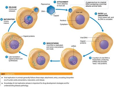
Multiplication of Animal Viruses
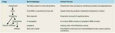
The multiplication of animal viruses involves several steps:
Attachment: Virus attaches to host cell membrane.
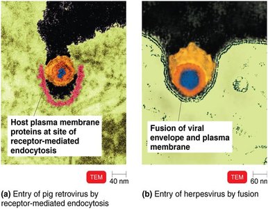
Entry: By receptor-mediated endocytosis or fusion.
Uncoating: Viral or host enzymes remove the capsid.
Biosynthesis: Production of viral nucleic acid and proteins.
Maturation: Assembly of viral components.
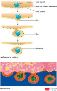
Release: By budding (enveloped viruses) or rupture (nonenveloped viruses).
Biosynthesis of DNA and RNA Viruses
DNA viruses: Replicate DNA in the nucleus using viral enzymes; synthesize capsid in the cytoplasm using host enzymes.
RNA viruses: Replicate in the cytoplasm using RNA-dependent RNA polymerase. Positive-sense RNA acts as mRNA; negative-sense RNA must be transcribed to positive-sense RNA first.
Classification of Viruses Affecting Humans
Viruses are classified by their nucleic acid type, structure, and replication strategy. The Baltimore classification scheme is commonly used. Major families include:
Class | Genome Type | Family | Example | Clinical Features |
|---|---|---|---|---|
I | dsDNA | Herpesviridae | Herpes simplex virus | Cold sores, chickenpox |
II | ssDNA | Parvoviridae | Parvovirus B19 | Fifth disease |
III | dsRNA | Reoviridae | Rotavirus | Gastroenteritis |
IV | +ssRNA | Picornaviridae | Poliovirus | Polio |
V | -ssRNA | Rhabdoviridae | Rabies virus | Rabies |
VI | ssRNA-RT | Retroviridae | HIV | AIDS |
VII | dsDNA-RT | Hepadnaviridae | Hepatitis B virus | Hepatitis B |
Viruses and Cancer
Oncogenes and Transformation
Some viruses can cause cancer by integrating their genetic material into the host genome, leading to transformation of normal cells into tumor cells. Oncogenes are genes that can induce uncontrolled cell growth. Oncogenic viruses may be DNA or RNA viruses (retroviruses).
DNA oncogenic viruses: Adenoviridae, Herpesviridae, Papovaviridae, Hepadnaviridae
RNA oncogenic viruses: Retroviridae (e.g., HTLV-1, HTLV-2)
Latent and Persistent Viral Infections
Viruses can establish different types of infections:
Latent infections: Virus remains dormant in host cells and may reactivate (e.g., herpes simplex virus, varicella-zoster virus).
Persistent infections: Virus is continuously present and released over a long period, often causing progressive disease (e.g., HIV, hepatitis B).
Viroids and Prions
Viroids
Viroids are infectious agents composed solely of short strands of circular, single-stranded RNA without a protein coat. They cause diseases in plants, such as potato spindle tuber disease.
Prions
Prions are infectious proteins that lack nucleic acids. They cause neurodegenerative diseases by inducing abnormal folding of normal cellular proteins, leading to brain damage. Examples include Creutzfeldt-Jakob disease and mad cow disease.
PrPC: Normal cellular prion protein.
PrPSc: Abnormal, disease-causing form that accumulates in brain tissue.
Additional info: The Baltimore classification scheme organizes viruses based on their genome type and replication strategy, which is crucial for understanding their biology and for developing antiviral therapies.
