 Indietro
Viruses, Viroids, and Prions: Structure, Replication, and Pathogenesis
Guida di studio - Note intelligenti
 Appunti personalizzati basati sui tuoi materiali, ampliati con definizioni chiave, esempi e contesto.
Viral Life Cycles and Bacteriophages
Lytic and Lysogenic Cycles of Bacteriophages
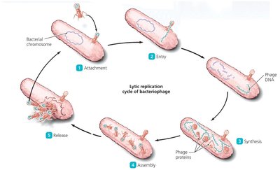
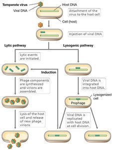
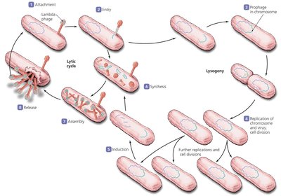
Bacteriophages (viruses that infect bacteria) can undergo two main types of replication cycles: the lytic cycle and the lysogenic cycle. These cycles determine how phages interact with their bacterial hosts and influence their use in biotechnology and medicine.
Lytic Cycle: The phage takes over the host cell machinery to produce new virions, ultimately lysing (destroying) the host cell to release progeny.
Lysogenic Cycle: The phage genome integrates into the host bacterial chromosome, becoming a prophage. The viral DNA is replicated along with the host genome and can remain dormant until induced to enter the lytic cycle.
Induction: Environmental signals can trigger the prophage to excise from the host genome and enter the lytic cycle.
Phage Therapy: Using Bacteriophages to Treat Bacterial Infections
Bacteriophages, especially lytic phages, are being explored as alternatives to antibiotics for treating bacterial infections. Their specificity for bacterial hosts allows targeted therapy, reducing harm to beneficial microbiota.
Lytic phages are preferred for therapy because they rapidly destroy bacterial cells.
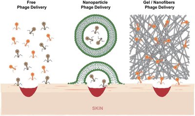
Phages can be delivered topically, orally, or via inhalation, depending on the infection site.
Genetic modification can enhance phage specificity and efficacy.
Animal Viruses: Life Cycles and Infection Types
Types of Viral Infections in Animal Cells
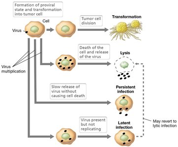
Animal viruses can cause several types of infections, each with distinct outcomes for the host cell:
Virulent (lytic) infection: Host cell is lysed to release new virions.
Persistent infection: Virions are released slowly without cell lysis.
Latent infection: Viral genome integrates into host DNA and remains dormant, similar to lysogeny in bacteria.
Transformation: Virus induces uncontrolled cell division, potentially leading to cancer.
Entry and Uncoating of Animal Viruses
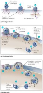
Animal viruses utilize specific mechanisms to enter host cells, often determined by the presence or absence of a viral envelope. Uncoating is essential for releasing the viral genome into the host cell.
Direct penetration: Capsid binds to the membrane, genome enters the cell, capsid remains outside.
Membrane fusion: Viral envelope fuses with the cell membrane, nucleocapsid enters the cytoplasm.
Endocytosis: Entire virion is engulfed by the cell, then uncoated inside.
Uncoating: Viral genome must be released from the capsid for replication and transcription.
Synthesis of Viral Components and Unique Viral Enzymes
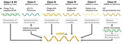
Viruses rely on host cell machinery for replication but may encode unique enzymes to facilitate their life cycle, especially RNA viruses.
Host enzymes: DNA polymerase (DNA replication), RNA polymerase (transcription).
Viral enzymes:
RNA-dependent RNA polymerase (RDRP): Synthesizes RNA from an RNA template (used by RNA viruses).
Reverse transcriptase (RNA-dependent DNA polymerase): Synthesizes DNA from an RNA template (used by retroviruses like HIV).
These enzymes are often packaged within the virion to ensure successful infection of new host cells.

Viral Budding and Virion Release
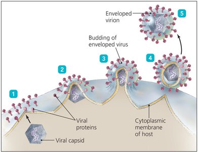
Enveloped animal viruses acquire their lipid envelope from host cell membranes during the budding process. This process allows virions to exit the cell without causing immediate lysis.
Budding can occur from the plasma membrane, endoplasmic reticulum, or nuclear membrane.
Viral proteins mediate membrane wrapping and release.
Some viruses require specific enzymes (e.g., neuraminidase in influenza) to complete budding.
Laboratory Methods: Plaque Assays
Quantifying Viruses Using Plaque Assays
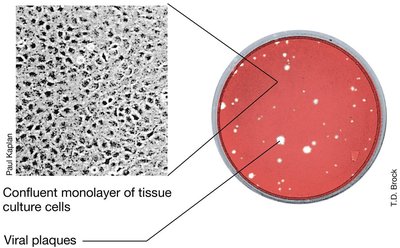
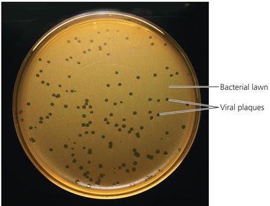
Plaque assays are used to determine the concentration of infectious virus particles in a sample. Plaques are clear zones formed by virus-induced cell lysis in a lawn of host cells.
Used for both bacteriophages (bacterial lawns) and animal viruses (tissue culture monolayers).
Results are expressed as plaque-forming units (PFU) per milliliter.
Acellular Infectious Agents: Viruses, Viroids, and Prions
Viroids: Infectious RNA Molecules
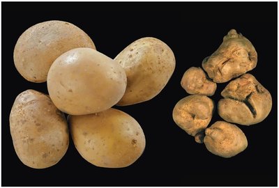
Viroids are the smallest known infectious agents, consisting solely of a short strand of circular, single-stranded RNA without a protein coat. They infect plants and can cause significant agricultural damage.
Do not encode proteins; replicate using host cell enzymes.
RNA folds into double-stranded regions for stability.
Entry into plant cells occurs through wounds, not receptor-mediated.

Viroid-Mediated Pathogenesis
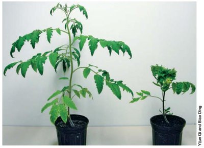
Viroid infection can lead to a range of symptoms in plants, from asymptomatic to severe growth defects, malformations, and death. Pathogenesis is often due to interference with host gene expression.
Viroids may cause degradation of specific plant mRNAs, inhibiting protein synthesis.
Only infect plants, not animals or microbes.
Prions: Infectious Proteins
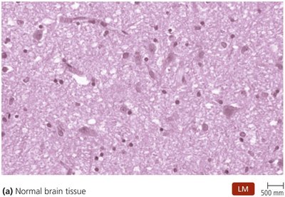
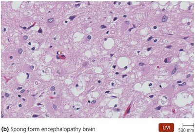
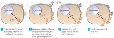
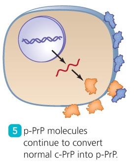
Prions are misfolded proteins that can induce normal proteins to adopt the pathogenic conformation, leading to neurodegenerative diseases known as transmissible spongiform encephalopathies (TSEs).
Contain no nucleic acid genome.
Diseases include Creutzfeldt-Jakob disease, kuru, fatal familial insomnia, bovine spongiform encephalopathy (mad cow disease), and chronic wasting disease in cervids.
Prion propagation depends on the ability of the misfolded protein (p-PrP) to convert normal protein (c-PrP) into the pathogenic form.
Prion diseases have long incubation periods and are highly resistant to standard decontamination methods.
Prion Destruction and Resistance
Prions are exceptionally resistant to conventional sterilization methods, including boiling, radiation, and standard autoclaving. Specialized protocols are required for decontamination.
CDC recommends immersion in 1N NaOH followed by autoclaving at high temperature.
Enzymatic treatments are being developed for prion decontamination.
Chronic Wasting Disease (CWD) in Cervids
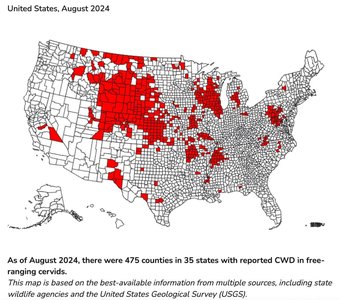
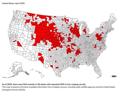
Chronic wasting disease is a prion disease affecting deer, elk, and related species. It is spreading across North America, with prions detectable in various tissues and bodily fluids.
As of April 2025, CWD was reported in 543 counties across 36 states in the U.S.

Comparison of Bacteria, Viruses, Viroids, and Prions
The following table summarizes the key differences among bacteria, viruses, viroids, and prions, focusing on structure, genetic material, and biological properties.
Bacteria | Viruses | Viroids | Prions | |
|---|---|---|---|---|
Width | 200–2000 nm | 10–400 nm | 2–10 nm | 5 nm |
Nucleic Acid | Both DNA and RNA | Either DNA or RNA, never both | RNA only | None |
Protein | Present | Present (capsid) | Absent | Present (PrP) |
Cytoplasmic Membrane | Present | Absent (some enveloped viruses have a membrane) | Absent | Absent |
Functional Ribosomes | Present | Absent | Absent | Absent |
Growth | Present | Absent | Absent | Absent |
Self-Replicating | Present | Some bacteriophages respond to a host cell by injecting their genomes | Absent | No; transforms PrP protein already present in cell |
Metabolism | Present | Absent | Absent | Absent |
Summary
This guide covers the structure, replication, and pathogenesis of viruses, viroids, and prions, highlighting their differences from cellular life forms and their significance in microbiology and medicine.
