 Indietro
Pathophysiology of Inflammation, Wound Healing, and Cancer: Study Notes for Personal Health
Guida di studio - Note intelligenti
 Appunti personalizzati basati sui tuoi materiali, ampliati con definizioni chiave, esempi e contesto.
Inflammation Fundamentals
Definition and Causes of Inflammation
Inflammation is a protective response triggered by injury, infection, or harmful stimuli. It involves coordinated cellular and vascular changes to identify, contain, and remove the cause, clear damaged tissue, and initiate repair. Both infectious and sterile injuries can provoke inflammation.
Biologic/Infection: Bacteria, viruses, and toxins activate immune defenses.
Physical Agents: Trauma, sunlight, and radiation cause direct tissue injury.
Chemical Stimuli: Chemicals and venoms damage cells or trigger immune reactions.
Reduced Oxygen: Anoxia, hypoxia, or ischemia injure cells due to inadequate oxygen delivery.
Sequence of the Inflammatory Response
The inflammatory response follows a predictable sequence:
Recognition: Detection of injury or infection.
Chemical Signaling: Release of mediators (e.g., histamine, prostaglandins).
Vasodilation: Increased blood flow causes redness and warmth.
Increased Permeability: Capillaries allow plasma proteins and leukocytes into tissue, causing swelling.
Cellular Cleanup: Neutrophils and macrophages remove debris and pathogens.
Resolution and Repair: Tissue regeneration or scar formation begins.
Classic Signs and Vascular Actions
Redness: Due to vasodilation and increased blood flow.
Heat: Local hyperemia brings warm blood to the area.
Swelling: Plasma and proteins leak into tissue.
Pain: Edema and mediators stimulate nerve endings.
Loss of Function: Pain and swelling limit movement.
Four Linked Actions in Inflammation
Hemodynamic Change: Capillary dilation increases warmth and redness.
Increased Permeability: Endothelial gaps allow fluid and cells to leave vessels, causing edema and pain.
Cell Migration: Leukocytes move to the site, sometimes forming pus.
Mediator/Hormonal Control: Chemical mediators coordinate and restrain the response.
Increased Capillary Permeability
Permeability refers to how easily substances pass through vessel walls. During inflammation, permeability increases, allowing fluid, proteins, and white blood cells to enter tissue. This results in swelling and exudate formation.
Example: A twisted ankle swells due to increased vessel permeability.
Stages of the Inflammatory Response
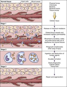
The inflammatory response after tissue injury occurs in three stages:
Stage 1: Injury triggers release of chemical mediators, vasodilation, and increased permeability. Proteins, water, and white blood cells move into tissue.
Stage 2: Phagocytic cells ingest foreign agents and debris, forming exudate.
Stage 3: Repair and regeneration restore tissue.
Cell Roles: Neutrophils and monocytes ingest bacteria and debris. Monocytes mature into macrophages for continued cleanup and repair coordination.
Important Terms
Leukocytosis: Increased white blood cell count supporting inflammation.
Phagocytosis: Engulfment and destruction of pathogens and debris.
Exudate: Protein-rich fluid and cells in inflamed tissue.
Resolution: Settling of the response after cause removal.
Chemical Mediators and Systemic Effects
Chemical mediators are emergency signals released by damaged cells and immune cells. They start, direct, and control inflammation.
Histamine: Vasodilation and increased permeability; released by mast cells.
Prostaglandins: Promote inflammation, pain, and fever.
Leukotrienes: Attract leukocytes and amplify response.
Bradykinin: Stimulates pain receptors.
Cytokines: Coordinate inflammation and immune activity; cause systemic symptoms.
Local vs Systemic Inflammation
Local: Redness, warmth, swelling, pain at injury site.
Systemic: Fever, headache, muscle aches, chills, sweating, fatigue, leukocytosis.
Systemic symptoms are caused by pyrogens and cytokines affecting body temperature and pain pathways.
Cortisol and Anti-inflammatory Therapy
Cortisol is an adrenal hormone that limits inflammation by reducing histamine release, stabilizing lysosomal membranes, and decreasing leukocyte influx.
Corticosteroid Medications: Mimic cortisol to suppress inflammation, but may increase infection risk and delay healing.
Example: Prolonged cortisone use can weaken immune responses and delay wound healing.
Wound Healing
Healing Process and Determinants
Repair and regeneration begin immediately after injury. Healing speed depends on tissue type, wound size, infection, age, health, perfusion, diabetes, and nutrition.
Macrophages: Remove debris and support repair.
Fibroblasts: Build collagen and elastin.
Granulation Tissue: New connective tissue and blood vessels.
Epithelial Cells: Cover and protect the wound surface.
Phases of Wound Healing
Inflammatory Phase: Hemostasis and cleanup.
Proliferative Phase: Tissue building and coverage.
Remodeling/Maturation Phase: Strengthening and scar formation.
Example: Healthy granulation tissue is pink-red and moist; yellow slough or green drainage requires assessment.
Nursing Applications
Begin discharge planning early.
Assess for infection, perfusion, nutrition, and wound care needs.
Coordinate with prescribers and dietitians for nutrition support.
Cancer and Neoplasms
Definition and Cell Control
Cancer is the presence of malignant cells that invade tissue and may spread. Normal cells control growth, function, and location; cancer develops when these controls are lost.
Benign Tumors: Localized, slower growth, resemble tissue of origin, do not invade or metastasize.
Malignant Tumors: Uncontrolled growth, abnormal appearance, loss of function, invasion, and metastasis.
Cancer Cell Characteristics
Abnormal appearance and function.
Uncontrolled division and invasion.
Production of tumor markers (e.g., CEA).
Ability to metastasize.
Tumor Markers and Metastasis
CEA: Monitors treatment and recurrence; not diagnostic alone.
Metastasis: Spread via blood, lymph, or direct invasion; common sites include liver, brain, bone, and lungs.
Malignant Transformation and Promoting Factors
Initiation: DNA damage by carcinogens.
Promotion: Growth-promoting influences accelerate cell division.
Progression: Tumor becomes invasive and accumulates mutations.
Metastasis: Cells spread and establish new colonies.
Classification, Differentiation, and Prognosis
Anatomic Site: Origin of tumor.
Stage: Extent of spread (TNM system).
Grade/Differentiation: How abnormal cells appear.
Prognosis: Estimated course based on type, stage, grade, and response.
TNM Staging System
Letter | Represents | Interpretation |
|---|---|---|
T | Primary tumor size/extent | Higher numbers = larger/more invasive tumor |
N | Regional lymph nodes | N0 = no nodes; higher numbers = more involvement |
M | Metastasis | M0 = no distant spread; M1 = distant metastasis present |
Warning Signs of Cancer
Change in bowel/bladder habits
Sore that does not heal
Unusual bleeding/discharge
Thickening/lump
Indigestion/difficulty swallowing
Change in wart/mole
Nagging cough/hoarseness
Persistent or unexplained changes should be assessed; early cancer may cause no symptoms.
Prevention and Early Detection
Balanced nutrition, healthy weight, physical activity, rest, and stress management.
Avoid known carcinogens (tobacco, harmful exposures).
Screening based on risk, age, and guidelines.
Cultural and Social Determinants
Consider access to care, discrimination, income, food access, health literacy, exposures, screening, and trust.
Use clear teaching, interpreters, referrals, and follow-up planning.
Integrated Review: Concept Links and Nursing Priorities
Vasodilation causes redness and heat; permeability causes swelling and pain.
Mediators drive local and systemic signs.
Corticosteroids reduce inflammation but delay healing.
Inflammation prepares for wound healing; normal-cell controls prevent cancer.
Loss of control leads to metastasis; stage and grade/differentiation affect prognosis.
Assess local vs systemic findings; monitor for infection.
Review corticosteroid use; support perfusion, glucose, nutrition, wound care, teaching, mobility, and discharge planning.
Report persistent cancer warning signs; support screening and referral.
Use precise, nonjudgmental communication; individualize oncology care.
Final memory line: INFLAMMATION responds. HEALING rebuilds. CORTISOL restrains. CANCER ignores control. METASTASIS spreads. SCREENING looks early.
