 Indietro
Biological Psychology: Structure and Function of the Brain
Guida di studio - Note intelligenti
 Appunti personalizzati basati sui tuoi materiali, ampliati con definizioni chiave, esempi e contesto.
Biological Psychology
Central Nervous System (CNS) and Peripheral Nervous System (PNS)
The nervous system is divided into the central and peripheral components, each with distinct roles in processing and transmitting information throughout the body.
Central Nervous System (CNS): Consists of the brain and spinal cord. Responsible for integrating sensory information and responding accordingly.
Peripheral Nervous System (PNS): Composed of nerves outside the CNS. Connects the CNS to limbs and organs, facilitating communication between the body and brain.
Anatomy & Function of the Brain
The brain is a complex organ with specialized regions responsible for various cognitive, sensory, and motor functions.
Cerebral Cortex: The outermost layer of the brain, characterized by folds (gyri) and grooves (sulci). Responsible for higher-order functions such as perception, reasoning, and voluntary movement.
Cerebral Hemispheres: The brain is divided into right and left hemispheres, separated by a deep groove called the longitudinal fissure.
Lateralization: Cognitive functions may rely more on one hemisphere than the other, a phenomenon known as lateralization.
Comparison of Hemispheric Functions
The left and right hemispheres of the brain specialize in different skills and actions.
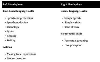
Left Hemisphere: Fine-tuned language skills (speech comprehension, production, reading, writing), making facial expressions, motion detection.
Right Hemisphere: Coarse language skills (simple speech, writing, tone of voice), visuospatial skills (perceptual grouping, face perception).
Lobes of the Brain
The cerebral cortex is divided into four main lobes, each with distinct functions:
Frontal Lobe: Responsible for executive functions, planning, decision-making, voluntary movement (motor cortex), and speech production (Broca's area).
Parietal Lobe: Processes sensory information (touch, pressure, pain, temperature), spatial perception, and guides attention.
Temporal Lobe: Involved in hearing, language comprehension (Wernicke's area), memory, and facial recognition (fusiform face area).
Occipital Lobe: Dedicated to visual processing.
Brain Structure and Connectivity
Gray Matter: Contains cell bodies and dendrites of neurons.
White Matter: Contains axons and myelinated sheaths, facilitating communication between brain regions.
Corpus Callosum: Large bundle of fibers connecting the two hemispheres, enabling interhemispheric communication.
Protective Membranes and Fluid
Meninges: Three protective layers (dura mater, arachnoid mater, pia mater) surrounding the brain and spinal cord.
Cerebrospinal Fluid (CSF): Surrounds the brain and spinal cord, filling ventricles and aqueducts, providing cushioning and protection.
Subcortical Structures
Subcortical regions play crucial roles in movement, emotion, and memory.
Basal Ganglia: Controls movement.
Limbic System: Includes the amygdala (emotional center), thalamus (sensory relay station), hypothalamus (regulates internal states and motivated behavior), and hippocampus (memory formation).
Cerebellum: Coordinates movement and assists in learning motor skills.
Brainstem: Composed of midbrain (visual and auditory tracking), pons (triggers dreams, connects to cerebellum), and medulla (regulates basic functions like heartbeat and breathing).
Neurons and Signal Transmission
Neurons are specialized cells for communication within the nervous system.
Subcomponents: Cell body (soma), nucleus, dendrites (receive signals), axon (sends signals).
Signal Transmission: Synaptic vesicles travel through the axon to axon terminals, where an action potential triggers the release of neurotransmitters into the synaptic gap. Neurotransmitters bind to receptor sites on dendrites of nearby neurons.
Reuptake: Recycling of neurotransmitters after signal transmission.
Neurotransmitters
Neurotransmitters are chemical messengers that facilitate communication between neurons.
Glutamate: Associated with enhanced learning and memory; excessive levels may contribute to disorders like schizophrenia.
GABA (gamma-aminobutyric acid): Inhibitory neurotransmitter; certain drugs bind to GABA to treat anxiety, insomnia, depression, and epilepsy.
Monoamines: Includes norepinephrine (motivation, reward), dopamine (motivation, reward), and serotonin (mood regulation).
Myelin Sheath and Signal Efficiency
Myelin Sheath: Insulates axons, speeding up electrical signal transmission.
Nodes of Ranvier: Gaps along axons that facilitate rapid signal conduction.
Oligodendrocytes: Cells that create myelin sheaths in the CNS.
Damage to Myelin: Disrupts signal efficiency, as seen in multiple sclerosis.
Neuroplasticity
The brain's ability to change and adapt in response to learning, experience, or injury.
Synaptogenesis: Formation of new synaptic connections.
Pruning: Elimination of excess neurons and axons to streamline neural organization.
Myelination: Insulation of axons by myelin sheaths, enhancing communication.
Structural Neuroplasticity: Changes in neuron shape or dendrite number.
Neuroimaging Techniques
Neuroimaging allows researchers to study brain structure and function using various technologies.
MRI (Magnetic Resonance Imaging): Non-invasive procedure for studying brain structure and function. Disadvantages include cost and limited accessibility.
PET (Positron Emission Tomography): Measures brain activity using radioactive substances. Disadvantage: invasive procedure.
Deep Brain Stimulation (DBS): Involves implanting electrodes to provide direct electrical stimulation.
Transcranial Magnetic Stimulation (TMS): Uses strong magnetic fields applied to the skull to enhance or interrupt brain function; non-invasive.
Summary Table: Hemispheric Functions
Left Hemisphere | Right Hemisphere |
|---|---|
Fine-tuned language skills:
Actions:
| Coarse language skills:
Visuospatial skills:
|
Key Equations
Action Potential: The change in electrical potential associated with the passage of an impulse along the membrane of a neuron.
Additional info: Some details were inferred based on standard introductory psychology content and context clues from the lecture slides.
