 Indietro
Neurophysiology: Structure and Function of Neurons and Glia
Guida di studio - Note intelligenti
 Appunti personalizzati basati sui tuoi materiali, ampliati con definizioni chiave, esempi e contesto.
Structure and Function of Neurons and Glia
Overview of Glial Cells
Glial cells are non-neuronal cells in the nervous system that provide support and protection for neurons. They play essential roles in maintaining homeostasis, forming myelin, and participating in signal transmission in the nervous system.
Macroglia: Includes astrocytes, ependymal cells, oligodendrocytes, and Schwann cells.
Microglia: Act as the immune cells of the central nervous system (CNS).
Astrocytes
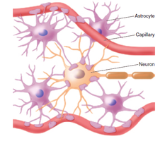
Astrocytes are star-shaped glial cells in the CNS that perform a variety of functions essential for neuronal health and signaling.
Nutrient Transfer: Facilitate the transfer of nutrients from blood vessels to neurons.
Blood-Brain Barrier: Block the movement of toxins into neural tissue.
Structural Support: Hold neurons in place and contribute to brain development.
Learning and Memory: Involved in synaptic plasticity and memory formation.
Debris Cleanup: Remove dead neurons and maintain extracellular environment.
Ependymal Cells
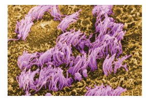
Ependymal cells line the ventricles of the brain and the central canal of the spinal cord, playing a key role in cerebrospinal fluid (CSF) dynamics.
CSF Production: Produce and regulate CSF in the choroid plexus.
CSF Circulation: Circulate CSF via cilia movement.
Protection: Act as a barrier against viruses entering the CNS.
Oligodendrocytes and Schwann Cells
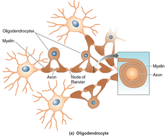
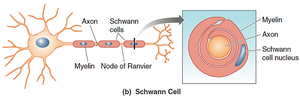
These glial cells are responsible for the formation and maintenance of the myelin sheath, which insulates axons and speeds up neural transmission.
Oligodendrocytes: Myelinate axons in the CNS; each cell can myelinate multiple axons.
Schwann Cells: Myelinate axons in the peripheral nervous system (PNS); each cell myelinates a single axon segment and aids in axonal regeneration.
Microglia
Microglia are the resident immune cells of the CNS, responsible for responding to injury and disease.
Phagocytosis: Clean up brain debris, including dead cells and synapses.
Synaptic Pruning: Remove unnecessary synapses during development and disease.
Dysregulation: Overactivation can contribute to neurodegenerative diseases such as Alzheimer's, Parkinson's, and multiple sclerosis.
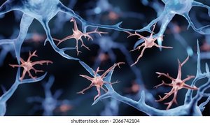
Structure of the Neuron
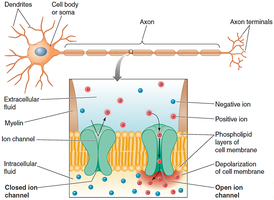
Cell Membrane
The neuron’s cell membrane is a phospholipid bilayer that separates the intracellular and extracellular environments, maintaining the ionic gradients necessary for electrical signaling.
Ion Pumps: Use energy (ATP) to move ions against their concentration gradients (e.g., sodium-potassium pump, calcium pump).
Ion Channels: Allow ions to move passively; can be voltage-gated or ligand-gated.
Cytoskeleton
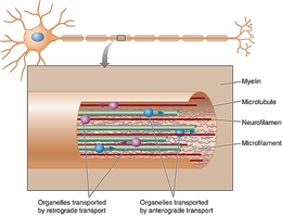
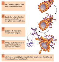
The cytoskeleton provides structural support and facilitates intracellular transport within neurons.
Intermediate Filaments (Neurofilaments): Provide structural stability.
Microfilaments: Involved in changing the shape and length of axons and dendrites.
Microtubules: Transport materials within the neuron via anterograde (cell body to periphery) and retrograde (periphery to cell body) transport.
Dendrites
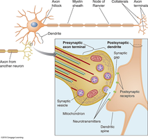
Dendrites are branched extensions of the neuron that receive synaptic inputs from other neurons.
Presynaptic Neuron: Sends the signal.
Postsynaptic Neuron: Receives the signal via dendrites.
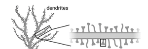
Dendritic Spines: Increase the surface area for synaptic contacts.
Axons
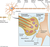
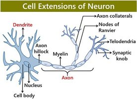
Axons are long projections that transmit electrical impulses away from the cell body toward other neurons or effectors.
Axon Hillock: Site where action potentials are initiated.
Myelin Sheath: Insulates the axon, increasing conduction speed.
Nodes of Ranvier: Gaps in myelin where action potentials are regenerated.
Axon Terminals: Release neurotransmitters to communicate with target cells.
White Matter: Composed of myelinated axons in the CNS.
Structural and Functional Variations of Neurons
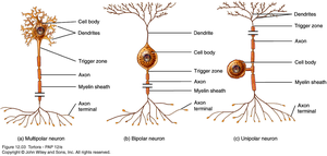
Neurons can be classified based on their structure and function.
Multipolar Neurons: Multiple dendrites, one axon; common in motor neurons.
Bipolar Neurons: One dendrite, one axon; found in sensory organs.
Unipolar Neurons: Single process that splits into two branches; common in sensory neurons.
Type | Structure | Function |
|---|---|---|
Motor Neuron | Multipolar | CNS to periphery |
Sensory Neuron | Unipolar/Bipolar | Periphery to CNS |
Interneuron | Multipolar/Bipolar | Bridge between sensory and motor systems |
Neuronal Signal Transmission
Overview of Signal Transmission
Neurons communicate via electrical and chemical signals. The 'decision' to fire an action potential depends on whether the membrane potential crosses a threshold voltage.
Electrical Signaling: Action potentials travel along the axon.
Chemical Signaling: Neurotransmitters are released at synapses to communicate with other neurons.
Synaptic Transmission
A synapse is the junction between two neurons, consisting of a presynaptic terminal, synaptic cleft, and postsynaptic membrane. There are two main types of synapses:
Electrical Synapses (Gap Junctions): Direct, rapid transmission via protein channels connecting adjacent neurons.
Chemical Synapses: Neurotransmitters are released from the presynaptic neuron and bind to receptors on the postsynaptic neuron.
Ion Channels and Receptors
Ion channels are proteins that allow specific ions to pass through the membrane, crucial for generating electrical signals.
Ionotropic Receptors: Ligand-gated, fast and direct action.
Metabotropic Receptors: G-protein coupled, slower and indirect, with longer-lasting effects.
Voltage-Gated Channels: Open in response to changes in membrane potential.
Excitatory and Inhibitory Postsynaptic Potentials (EPSPs and IPSPs)
Postsynaptic potentials are changes in membrane potential due to synaptic activity.
EPSP: Temporary depolarization caused by the influx of positive ions, making the neuron more likely to fire an action potential.
IPSP: Temporary hyperpolarization caused by the influx of negative ions, making the neuron less likely to fire.
Summation of Signals
Neurons integrate multiple EPSPs and IPSPs through spatial and temporal summation. If the net effect at the axon hillock reaches the threshold, an action potential is triggered.
Spatial Summation: Multiple inputs from different locations.
Temporal Summation: Multiple inputs from the same location in rapid succession.
Action Potential Generation and Propagation
An action potential is an all-or-none electrical impulse generated at the axon initial segment when the threshold is reached.
Initiation: Occurs at the axon hillock/initial segment.
Propagation: Action potential travels along the axon, with myelination increasing speed via saltatory conduction at the nodes of Ranvier.
Refractory Periods: Absolute (no new AP possible) and relative (requires stronger stimulus).
Termination of Synaptic Transmission
Neurotransmitter action is terminated by:
Diffusion: Neurotransmitter molecules diffuse away from the synaptic cleft.
Enzymatic Degradation: Enzymes break down neurotransmitters.
Reuptake: Neurotransmitters are reabsorbed by the presynaptic neuron (e.g., SSRIs block serotonin reuptake).
Summary Table: Glial Cell Types and Functions
Glial Cell Type | Location | Main Function |
|---|---|---|
Astrocyte | CNS | Support, nutrient transfer, blood-brain barrier, debris cleanup |
Ependymal Cell | CNS | CSF production and circulation |
Oligodendrocyte | CNS | Myelination of axons |
Schwann Cell | PNS | Myelination and axon regeneration |
Microglia | CNS | Immune defense, debris cleanup |
