 Indietro
Blood Vessels and Circulatory Physiology: Study Notes
Guida di studio - Note intelligenti
 Appunti personalizzati basati sui tuoi materiali, ampliati con definizioni chiave, esempi e contesto.
Blood Vessels: Structure and Function
Major Arteries of the Body
The human body contains numerous arteries that supply oxygenated blood to various tissues. Key arteries include the superficial temporal artery, facial artery, common carotid artery, brachial artery, radial artery, femoral artery, popliteal artery, posterior tibial artery, and dorsalis pedis artery. These arteries are essential for maintaining tissue perfusion and can be used as pressure points to control bleeding.
Monitoring Circulatory Efficiency
Vital signs such as pulse, blood pressure, respiratory rate, and body temperature are used to assess circulatory efficiency. The pulse is a pressure wave caused by the expansion and recoil of arteries, commonly measured at the radial artery. Pressure points are locations where arteries are close to the body surface and can be compressed to stop blood flow.
Measuring Blood Pressure
Blood pressure is measured indirectly using the auscultatory method with a sphygmomanometer. The cuff is inflated above systolic pressure and then slowly released while listening for Korotkoff sounds with a stethoscope. Systolic pressure (normally <120 mm Hg) is the pressure when sounds first occur, and diastolic pressure (normally <80 mm Hg) is when sounds disappear.
Variations and Alterations in Blood Pressure
Blood pressure varies with age, sex, weight, race, mood, and posture. Hypertension is defined as sustained elevated arterial pressure of 140/90 mm Hg or higher. Prehypertension refers to values elevated but not yet in the hypertension range. Hypotension is blood pressure below 90/60 mm Hg and is usually not a concern unless it leads to inadequate tissue perfusion.
Homeostatic Imbalances: Hypertension and Hypotension
Prolonged hypertension is a major cause of heart failure, vascular disease, renal failure, and stroke. Primary hypertension has no identifiable cause and is influenced by heredity, diet, obesity, age, diabetes, stress, and smoking. Secondary hypertension is due to identifiable disorders such as kidney disease or endocrine disorders. Orthostatic hypotension is temporary low BP upon standing, while chronic hypotension may indicate poor nutrition or endocrine disorders. Acute hypotension is a sign of circulatory shock.
Blood Flow Through Body Tissues
Tissue Perfusion
Tissue perfusion is essential for delivering oxygen and nutrients, removing wastes, gas exchange in the lungs, absorption of nutrients in the digestive tract, and urine formation in the kidneys. The rate of blood flow must be precisely regulated to meet tissue needs.
Distribution of Blood Flow
Blood flow distribution changes between rest and exercise. During strenuous exercise, blood flow to skeletal muscles increases significantly, while flow to other organs may decrease.
Organ | Blood Flow at Rest (ml/min) | Blood Flow During Exercise (ml/min) |
|---|---|---|
Brain | 750 | 750 |
Heart | 250 | 750 |
Skeletal Muscles | 1200 | 12,500 |
Skin | 500 | 1900 |
Kidneys | 1100 | 600 |
Abdomen | 1400 | 600 |
Other | 600 | 400 |
Total | 5800 | 17,500 |
Velocity of Blood Flow
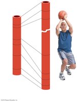
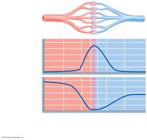
The velocity of blood flow changes as it travels through the systemic circulation. It is fastest in the aorta, slowest in capillaries, and increases in veins. This is inversely related to the total cross-sectional area of the vessels. Slow capillary flow allows adequate time for exchange between blood and tissues.
Autoregulation of Blood Flow
Intrinsic and Extrinsic Controls
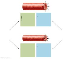
Autoregulation is the automatic adjustment of blood flow to each tissue based on its needs. It is controlled intrinsically by modifying the diameter of local arterioles and is independent of mean arterial pressure (MAP). Organs regulate their own blood flow by varying resistance in their arterioles.
Types of Autoregulation
Metabolic controls: Vasodilation occurs in response to declining tissue O2 and substances from metabolically active tissues (e.g., H+, K+, adenosine, prostaglandins).
Myogenic controls: Vascular smooth muscle responds to stretch; increased pressure promotes vasoconstriction, reduced stretch promotes vasodilation.
Long-term Autoregulation
When short-term autoregulation cannot meet tissue nutrient requirements, angiogenesis occurs, increasing the number of vessels to a region and enlarging existing vessels. This is common in the heart when coronary vessels are occluded or in people living at high altitudes.
Blood Flow in Specific Organs
Skeletal Muscles
Blood flow varies with muscle fiber type and activity. At rest, myogenic and neural mechanisms predominate. During activity, blood flow increases in proportion to metabolic activity, and local controls override sympathetic vasoconstriction.
Brain
Blood flow to the brain is constant due to neurons' intolerance of ischemia. Metabolic controls (decreased pH or increased CO2) cause vasodilation. Myogenic controls respond to changes in MAP. Extreme systemic pressure changes can cause syncope or cerebral edema.
Skin
Blood flow through the skin supplies nutrients, regulates body temperature, and provides a blood reservoir. Regulation is primarily neural.
Lungs
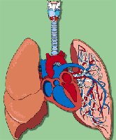
The pulmonary circuit is unique, with low resistance and pressure. Autoregulatory mechanisms are opposite to most tissues: low O2 causes vasoconstriction, high O2 promotes vasodilation, directing blood flow to O2-rich areas.
Heart
During ventricular systole, coronary vessels are compressed and blood flow ceases; myoglobin supplies O2. During diastole, high aortic pressure forces blood through coronary circulation. During exercise, coronary vessels dilate, increasing blood flow three to four times.
Capillary Exchange and Fluid Movements
Capillary Exchange of Gases and Nutrients
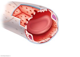
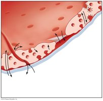
Exchange occurs by diffusion down concentration gradients. Lipid-soluble molecules diffuse through endothelium, water-soluble solutes pass through clefts and fenestrations, and larger molecules are transported via pinocytotic vesicles or caveolae.
Bulk Flow: Fluid Movements
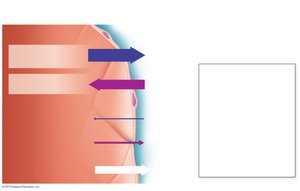
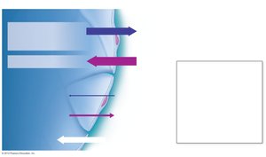
Fluid leaves capillaries at the arterial end and returns at the venous end. The direction and amount of fluid flow depend on hydrostatic and colloid osmotic pressures.
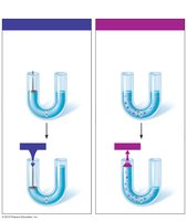
Hydrostatic pressure (HP): Pushes fluid across the boundary, due to blood pressure.
Osmotic pressure (OP): Pulls fluid across the boundary, due to plasma proteins.
Hydrostatic and Osmotic Pressure Interactions
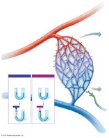
Capillary hydrostatic pressure (HPc) is higher at the arterial end (35 mm Hg) than at the venule end (17 mm Hg). Capillary colloid osmotic pressure (OPc) is ~26 mm Hg. Net filtration pressure (NFP) determines fluid movement:
Formula:
Net Filtration and Reabsorption
At the arteriolar end, net filtration occurs (NFP = 10 mm Hg), moving fluid out of the capillary. At the venous end, net reabsorption occurs (NFP = -8 mm Hg), moving fluid into the capillary.
Circulatory Shock
Types and Causes
Circulatory shock occurs when blood vessels are inadequately filled and blood cannot circulate normally, resulting in inadequate tissue perfusion. Types include:
Hypovolemic shock: Due to large-scale blood loss.
Vascular shock: Due to extreme vasodilation and decreased peripheral resistance.
Cardiogenic shock: Due to inefficient heart function.
Physiological Responses and Signs
Shock leads to inadequate tissue perfusion, anaerobic metabolism, movement of interstitial fluid into blood, and various compensatory mechanisms (activation of chemoreceptors, baroreceptors, hypothalamus, respiratory centers, and sympathetic nervous system). Signs include tachycardia, weak pulse, cold and clammy skin, reduced urine output, thirst, restlessness, and coma.
Example: Acute bleeding can trigger hypovolemic shock, leading to compensatory mechanisms to maintain blood pressure and perfusion.
