 Indietro
Bone Development, Growth, Remodeling, and Disorders
Guida di studio - Note intelligenti
 Appunti personalizzati basati sui tuoi materiali, ampliati con definizioni chiave, esempi e contesto.
Bone Development (Ossification)
Overview of Ossification
Ossification, also known as osteogenesis, is the process by which bone tissue forms. This process begins in the second month of embryonic development and continues throughout life as bones grow and remodel. There are two primary methods of bone development: endochondral ossification and intramembranous ossification.
Endochondral ossification: Bone forms by replacing hyaline cartilage. Most bones of the skeleton are formed this way.
Intramembranous ossification: Bone develops directly from mesenchymal tissue. This process forms flat bones such as those of the skull and clavicle.
Bone remodeling and repair are lifelong processes.
Endochondral Ossification
Process and Significance
Endochondral ossification is responsible for the formation of most bones, especially long bones. It involves the replacement of a hyaline cartilage model with bone tissue.
Step 1: Bone collar forms around the diaphysis of the hyaline cartilage model.
Step 2: Cartilage in the center of the diaphysis calcifies and develops cavities.
Step 3: The periosteal bud invades the internal cavities, and spongy bone forms.
Step 4: The diaphysis elongates, and a medullary cavity forms. Secondary ossification centers appear in the epiphyses.
Step 5: The epiphyses ossify. Hyaline cartilage remains only in the epiphyseal plates and articular cartilages.
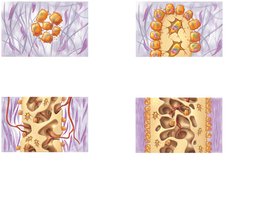
Additional info: The primary ossification center is typically located in the center of the diaphysis, while secondary centers develop in the epiphyses.
Intramembranous Ossification
Formation of Flat Bones
Intramembranous ossification forms flat bones such as the frontal, parietal, occipital, temporal bones, and the clavicle. This process begins with mesenchymal cells differentiating into osteoblasts within a fibrous connective tissue membrane.
Step 1: Ossification centers appear in the fibrous connective tissue membrane.
Step 2: Osteoid is secreted and calcifies, trapping osteoblasts which become osteocytes.
Step 3: Woven bone and periosteum form as osteoid accumulates between embryonic blood vessels.
Step 4: Lamellar bone replaces woven bone, and red marrow appears within the spongy bone (diploë).
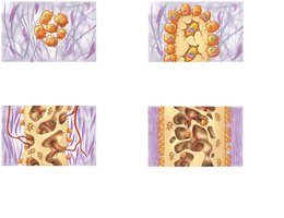
Additional info: This process is essential for the development of the cranial bones and clavicles.
Postnatal Bone Growth
Growth in Length (Interstitial Growth)
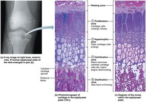
Long bones grow in length at the epiphyseal plate through interstitial growth. This process continues until adolescence, after which the epiphyseal plates close and are replaced by the epiphyseal line.
Growth stops at approximately 18 years in females and 21 years in males.
The epiphyseal plate consists of zones: resting, proliferation, hypertrophic, calcification, and ossification.
Growth in Width (Appositional Growth)
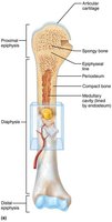
Bones increase in thickness through appositional growth, which involves the addition of new bone tissue by osteoblasts beneath the periosteum. This process is influenced by mechanical stress and continues throughout life.
Osteoblasts add bone matrix to the external surface, while osteoclasts remove bone from the internal surface.
Bones thicken in response to increased stress from muscle activity or weight-bearing.
Disorders of Bone Growth
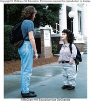
Dwarfism
Dwarfism refers to conditions resulting in short stature due to abnormal bone growth. Two main types are:
Achondroplastic dwarfism: Caused by failure of cartilage growth in the metaphysis, often due to a spontaneous mutation.
Pituitary dwarfism: Results from a lack of growth hormone, leading to normal body proportions but short stature.
Bone Remodeling
Continuous Bone Turnover
Bone remodeling is a dynamic process involving bone deposit by osteoblasts and bone resorption by osteoclasts. Approximately 5-7% of bone mass is recycled weekly.
Spongy bone is replaced every 3-4 years; compact bone every 10 years.
Bone deposit involves the secretion of osteoid and subsequent calcification.
Bone resorption involves osteoclasts breaking down bone matrix, releasing minerals into the blood.
Calcium Homeostasis and Hormonal Control
Regulation of Blood Calcium Levels
Calcium homeostasis is tightly regulated by hormonal control, primarily involving parathyroid hormone (PTH) and calcitonin.
PTH: Released by the parathyroid glands in response to low blood calcium levels; stimulates osteoclasts to resorb bone and release calcium into the blood.
Calcitonin: Released by the thyroid gland in response to high blood calcium levels; stimulates osteoblasts to deposit calcium into bone.
Normal blood calcium levels: 9–11 mg/100 ml.
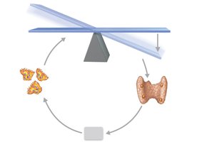
Additional info: Imbalances can lead to hypocalcemia (hyperexcitability) or hypercalcemia (nonresponsiveness).
Mechanical Stress and Bone Remodeling
Wolff’s Law
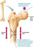
Wolff’s Law states that bones grow or remodel in response to the mechanical stresses placed upon them. This explains differences in bone density and structure based on activity level and handedness.
Bones are thicker where muscles attach and in dominant limbs.
Weight lifters have denser bones; bedridden individuals experience bone loss.
Fractures and Bone Repair
Classification of Fractures
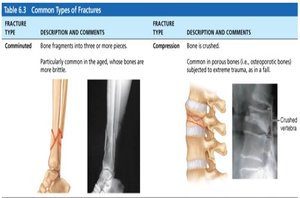
Fractures are classified by the position of bone ends, completeness of the break, and whether the skin is penetrated. Common types include:
Type | Description |
|---|---|
Comminuted | Bone fragments into three or more pieces; common in aged, brittle bones. |
Compression | Bone is crushed; common in porous bones subjected to trauma. |
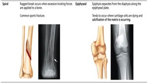
Spiral | Ragged break due to excessive twisting forces; common sports fracture. |
Epiphyseal | Epiphysis separates from diaphysis along epiphyseal plate; occurs where cartilage cells are dying. |
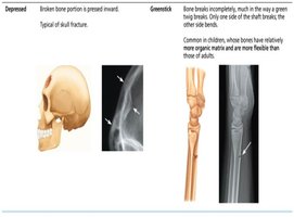
Depressed | Broken bone portion is pressed inward; typical of skull fracture. |
Greenstick | Bone breaks incompletely; common in children. |
Stages of Bone Healing
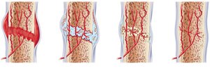
Bone healing occurs in four main stages:
Hematoma formation
Fibrocartilaginous callus formation
Bony callus formation
Bone remodeling
Osteoporosis
Pathophysiology and Risk Factors
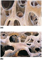
Osteoporosis is a condition characterized by severe loss of bone density, where bone resorption outpaces bone deposit. This leads to fragile bones and increased fracture risk.
Most common in postmenopausal women (30% by age 60-70, 70% by age 80).
Risk factors include insufficient exercise, poor diet (low calcium/protein), smoking, genetics, hormone imbalances, certain diseases (e.g., hyperthyroidism, diabetes), and alcohol or medication use.
Additional info: Prevention includes weight-bearing exercise, adequate calcium and vitamin D intake, and avoiding smoking and excessive alcohol.
