Indietro
Bone Development, Growth, Remodeling, and Fractures
Guida di studio - Note intelligenti
 Appunti personalizzati basati sui tuoi materiali, ampliati con definizioni chiave, esempi e contesto.
Bones and Skeletal Tissue: Bone Development and Growth
Ossification (Osteogenesis)
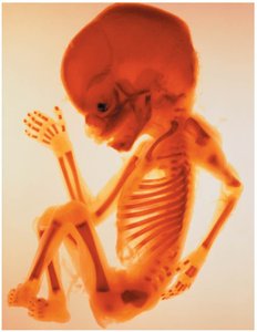
Ossification is the process of bone formation, beginning in the second month of embryonic development and continuing throughout life as bones grow, remodel, and repair. There are two primary methods of bone development: endochondral ossification and intramembranous ossification.
Endochondral Ossification: Bone forms by replacing hyaline cartilage. This process forms most bones of the skeleton, especially long bones.
Intramembranous Ossification: Bone develops directly from mesenchymal tissue. This process forms flat bones such as the frontal, parietal, occipital, temporal bones, and the clavicle.
Remodeling and repair of bone are lifelong processes, ensuring bone strength and calcium homeostasis.
Endochondral Ossification
Endochondral ossification is the process by which most bones are formed, replacing a hyaline cartilage model with bone tissue. The process occurs in several steps:
Bone collar forms around the diaphysis of the hyaline cartilage model.
Cartilage in the center of the diaphysis calcifies and develops cavities.
The periosteal bud invades the internal cavities, and spongy bone forms.
The diaphysis elongates, and a medullary cavity forms. Secondary ossification centers appear in the epiphyses.
The epiphyses ossify. Hyaline cartilage remains only in the epiphyseal plates and articular cartilages.
Intramembranous Ossification
Intramembranous ossification forms bone directly from mesenchymal tissue, without a cartilage precursor. This process is responsible for the formation of flat bones of the skull and the clavicle.
Ossification centers appear in the fibrous connective tissue membrane as mesenchymal cells differentiate into osteoblasts.
Osteoid is secreted and calcifies, trapping osteoblasts, which become osteocytes.
Woven bone and periosteum form as osteoid accumulates between embryonic blood vessels.
Lamellar bone replaces woven bone, and red marrow appears within the spongy bone (diploë).
Postnatal Bone Growth
Interstitial Growth (Lengthening of Long Bones)
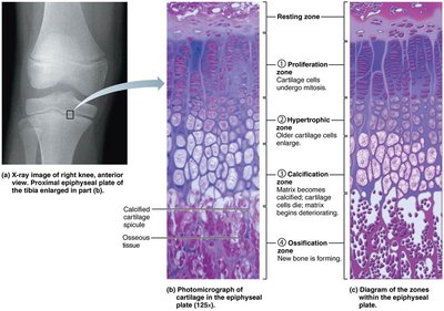
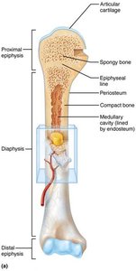
Long bones grow in length by interstitial growth at the epiphyseal plate, a layer of hyaline cartilage between the epiphysis and diaphysis. This process continues until the epiphyseal plates close, forming the epiphyseal line (around age 18 in females and 21 in males).
Zones of the Epiphyseal Plate:
Resting zone
Proliferation zone (cartilage cells undergo mitosis)
Hypertrophic zone (older cartilage cells enlarge)
Calcification zone (matrix calcifies, cartilage cells die)
Ossification zone (new bone forms)
Appositional Growth (Bone Thickening)
Bones increase in thickness (diameter) through appositional growth, which occurs as osteoblasts in the periosteum secrete new bone matrix on the external surface, while osteoclasts in the endosteum remove bone from the internal surface. This process allows bones to thicken in response to increased stress.
Growth Disorders
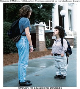
Achondroplastic Dwarfism: Caused by failure of cartilage growth in the metaphysis, often due to a spontaneous mutation. Results in short stature with abnormal proportions.
Pituitary Dwarfism: Caused by a lack of growth hormone, resulting in normal body proportions but short stature.
Bone Remodeling
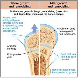
Bone remodeling is a continuous process where old bone is replaced by new bone tissue. About 5-7% of bone mass is recycled weekly. Spongy bone is replaced every 3-4 years, and compact bone every 10 years.
Bone Deposit: Osteoblasts secrete osteoid, which then calcifies.
Bone Resorption: Osteoclasts break down bone matrix, releasing minerals into the blood.
Control of Bone Remodeling
Hormonal Regulation
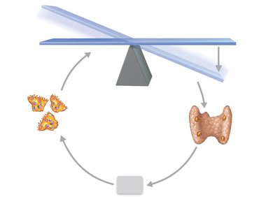
Bone remodeling is regulated by hormonal controls and mechanical stress. The main hormones involved are parathyroid hormone (PTH) and calcitonin.
Parathyroid Hormone (PTH): Released in response to low blood calcium levels. Stimulates osteoclasts to resorb bone, releasing calcium into the blood.
Calcitonin: Released in response to high blood calcium levels. Stimulates osteoblasts to deposit bone, lowering blood calcium.
Homeostatic Imbalances
Hypocalcemia: Low blood calcium, leading to muscle hyperexcitability and possible tetany.
Hypercalcemia: High blood calcium, causing muscle sluggishness and nonresponsiveness.
Mechanical Stress and Wolff's Law
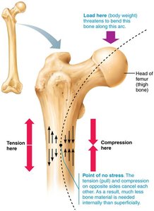
According to Wolff's Law, bones grow and remodel in response to the mechanical stresses placed upon them. This explains why dominant limbs are often thicker and why weight-bearing exercise strengthens bones.
Bone Fractures and Repair
Types of Fractures
Fractures are classified by the position of bone ends, completeness of the break, and whether the skin is penetrated. Common types include:
Fracture Type | Description and Comments |
|---|---|
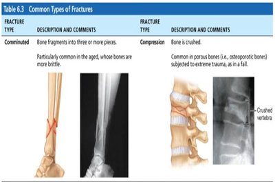
Comminuted | Bone fragments into three or more pieces; common in aged, brittle bones. |
Compression | Bone is crushed; common in porous bones subjected to trauma. |
Fracture Type | Description and Comments |
|---|---|
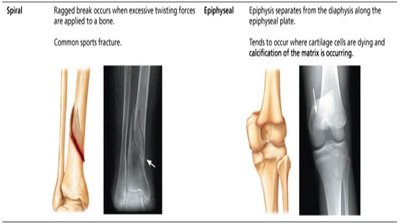
Spiral | Ragged break from excessive twisting; common sports fracture. |
Epiphyseal | Epiphysis separates from diaphysis at the epiphyseal plate; occurs where cartilage cells are dying. |
Fracture Type | Description and Comments |
|---|---|
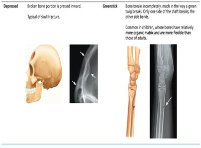
Depressed | Broken bone portion pressed inward; typical of skull fracture. |
Greenstick | Bone breaks incompletely, like a green twig; common in children. |
Healing of Bone Fractures
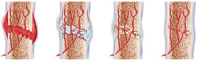
Bone repair occurs in four stages:
Hematoma forms (blood clot at fracture site).
Fibrocartilaginous callus forms (soft callus of cartilage and fibrous tissue).
Bony callus forms (spongy bone replaces soft callus).
Bone remodeling restores bone to original shape.
Osteoporosis
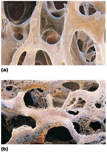
Osteoporosis is a condition characterized by severe loss of bone density, where bone resorption outpaces bone deposit. It is most common in postmenopausal women but can affect anyone with risk factors such as insufficient exercise, poor diet, smoking, genetics, or hormone-related conditions.
Normal bone: Dense with a regular structure.
Osteoporotic bone: Porous and fragile, increasing fracture risk.