 Indietro
Bones and Bone Structure: Study Notes for Anatomy & Physiology
Guida di studio - Note intelligenti
 Appunti personalizzati basati sui tuoi materiali, ampliati con definizioni chiave, esempi e contesto.
Chapter 6: Bones and Bone Structure
Introduction
This chapter explores the structure, classification, and functions of bones, as well as the cellular and molecular composition of bone tissue. Understanding these concepts is essential for comprehending the skeletal system's role in human anatomy and physiology.
Functions of the Skeletal System
Major Functions
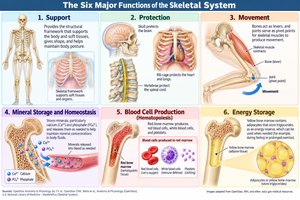
The skeletal system performs several critical functions necessary for survival and homeostasis:
Support: Provides the structural framework for the body, supporting soft tissues and giving shape to the body.
Protection: Shields vital organs such as the brain (protected by the skull), heart, and lungs (protected by the rib cage).
Movement: Serves as levers for muscles to act upon, enabling movement at joints.
Mineral Storage and Homeostasis: Stores minerals, primarily calcium and phosphate, and releases them into the bloodstream as needed.
Blood Cell Production: Houses red bone marrow, which produces red blood cells, white blood cells, and platelets (a process called hematopoiesis).
Energy Storage: Stores lipids in yellow bone marrow, serving as an energy reserve.
Composition of the Skeletal System
Overview
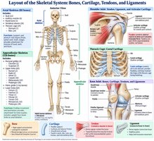
The skeletal system consists of bones, cartilage, tendons, and ligaments. Bones are the primary organs, while cartilage provides flexible support, and tendons and ligaments connect muscles to bones and bones to each other, respectively.
Classification of Bones by Shape
Types of Bones
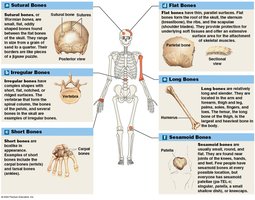
Bones are classified according to their shapes, which relate to their functions:
Sutural Bones: Small, irregular bones found between the flat bones of the skull.
Irregular Bones: Complex shapes, such as vertebrae and some skull bones.
Short Bones: Small and boxy, found in the wrist (carpals) and ankle (tarsals).
Flat Bones: Thin, parallel surfaces; examples include the sternum, ribs, and scapulae.
Long Bones: Relatively long and slender, such as the femur, humerus, and phalanges.
Sesamoid Bones: Usually small, round, and flat; develop inside tendons, e.g., the patella.
Classification of Bone Markings
Bone Markings and Their Functional Groups
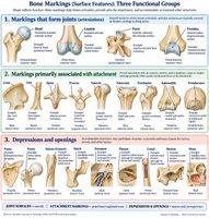
Bone markings are surface features that serve as sites for muscle, tendon, and ligament attachment, or as passages for nerves and blood vessels. They are grouped functionally as follows:
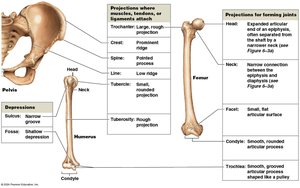
Markings that form joints: Head, condyle, facet, trochlea – smooth surfaces where bones articulate.
Markings primarily associated with attachment: Crest, line, spine, process, tubercle, tuberosity, trochanter, epicondyle, protuberance – provide attachment sites for tendons, muscles, and/or ligaments. Larger and rougher markings reflect greater mechanical forces.
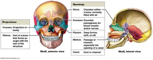
Depressions and openings: Fossa, fovea, sulcus/groove, notch, foramen, fissure, canal, meatus, sinus – accommodate other structures, participate in joints, or provide pathways for nerves and vessels.
Examples of Bone Markings
Specific examples of bone markings include:
Foramen: Opening for nerves and vessels.
Groove (sulcus): Furrow that protects vessels or nerves.
Process: Projection or bump for muscle attachment.
Sinus: Chamber within a bone, normally filled with air.
General Bone Structure
Long Bone Anatomy
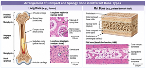
Long bones have a characteristic structure with distinct regions:
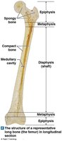
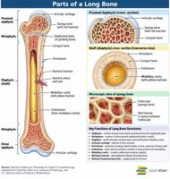
Diaphysis: The shaft, composed mainly of compact bone.
Epiphysis: The expanded ends, consisting mostly of spongy bone covered by compact bone.
Metaphysis: The region where the diaphysis and epiphysis meet; contains the growth plate in children.
Medullary Cavity: Central cavity containing bone marrow.
Compact vs. Spongy Osseous Tissue
Structural Differences
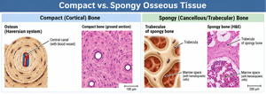
Both compact and spongy bone contain the same types of cells and mineralized matrix, but differ in organization:
Compact Bone (Cortical Bone): Densely packed; organized into osteons (Haversian systems) for strength and resistance to stress.
Spongy Bone (Cancellous/Trabecular Bone): Forms a network of trabeculae; lighter and supports bone marrow.
Bone Tissue (Osseous Tissue)
Composition and Characteristics
Bone tissue is a specialized connective tissue with a dense matrix and specialized cells:
Matrix: Two-thirds is calcium phosphate (as hydroxyapatite crystals), providing hardness and resistance to compression. One-third is collagen fibers, providing flexibility and tensile strength.
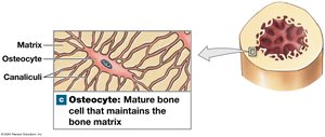
Osteocytes: Mature bone cells located in lacunae, connected by canaliculi for nutrient and waste exchange.
Periosteum: A membrane covering the outer surface of bones (except at joints), consisting of an outer fibrous layer and an inner cellular layer.
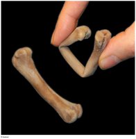
A bone lacking a calcified matrix appears normal but is very flexible, highlighting the importance of mineralization for bone strength.
Bone Cells
Types and Functions
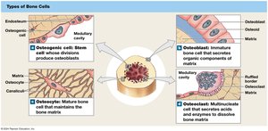
Bone contains four main types of cells, each with distinct roles:
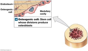
Osteogenic (Osteoprogenitor) Cells: Stem cells that divide to produce osteoblasts; found in the periosteum and endosteum; essential for fracture repair.
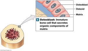
Osteoblasts: Immature bone cells responsible for osteogenesis (formation of new bone matrix); secrete osteoid and promote mineralization; become osteocytes when surrounded by matrix.
Osteocytes: Mature bone cells in lacunae; maintain bone matrix and help repair damaged bone; do not divide.
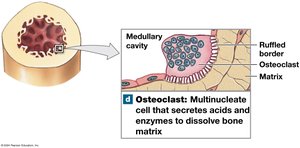
Osteoclasts: Large, multinucleate cells that resorb bone matrix (osteolysis); secrete acids and enzymes to dissolve bone and release minerals; not related to other bone cell lineages.
Cell Type | Main Function | Location |
|---|---|---|
Osteogenic Cell | Stem cell; produces osteoblasts | Periosteum, endosteum |
Osteoblast | Builds bone matrix (osteogenesis) | Bone surfaces |
Osteocyte | Maintains bone matrix | Lacunae within matrix |
Osteoclast | Resorbs bone matrix (osteolysis) | Bone surfaces |
Bone Matrix
Composition
Inorganic Component: Calcium phosphate (hydroxyapatite) and other salts (calcium carbonate, sodium, magnesium, fluoride) provide hardness and resistance to compression.
Organic Component: Collagen fibers form a flexible framework for mineral deposition, providing tensile strength and flexibility.
The balance between mineral and collagen components gives bone its unique combination of strength and flexibility.
Summary Table: Bone Tissue Components
Component | Proportion | Function |
|---|---|---|
Calcium Phosphate (Hydroxyapatite) | ~2/3 | Hardness, resistance to compression |
Collagen Fibers | ~1/3 | Flexibility, tensile strength |
Bone Cells | ~2% | Growth, maintenance, remodeling |
Additional info: The process of bone formation is called ossification, and bone remodeling is a continuous process involving both osteoblasts and osteoclasts to maintain bone health and adapt to stress.
