 Indietro
Cell Membrane Structure, Function, and Cellular Junctions
Guida di studio - Note intelligenti
 Appunti personalizzati basati sui tuoi materiali, ampliati con definizioni chiave, esempi e contesto.
Ch. 3 Cells: Structure and Function of the Cell Membrane
Overview of Levels of Organization
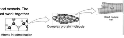
The human body is organized into hierarchical levels, each building upon the previous. Understanding these levels is essential for grasping how cells and their membranes contribute to overall physiology.
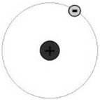
Atoms: The smallest units of matter, forming the basis of all substances.
Small Molecules: Combinations of atoms, such as water (H2O).
Macromolecules: Large, complex molecules like proteins and nucleic acids.
Cells: The basic structural and functional units of life.
Tissues: Groups of similar cells performing a common function.
Organs: Structures composed of two or more tissue types working together for specific functions.
Organ Systems: Groups of organs that cooperate to accomplish a common purpose.
Organism: The complete living being.
Structure and Function of the Cell Membrane
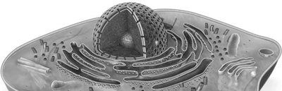
The cell membrane (also called the plasma membrane) is a dynamic structure that separates the interior of the cell from its external environment. It plays a critical role in maintaining cellular integrity and mediating interactions with the environment.
Separation of Compartments: Divides intracellular fluid from extracellular fluid.
Regulation of Transport: Controls the entry and exit of substances, maintaining homeostasis.
Cell Communication: Displays and secretes biological markers for cell recognition and signaling.
Fluid Mosaic Model of the Cell Membrane
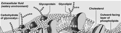
The cell membrane is described by the fluid mosaic model, which highlights its flexible, dynamic nature and the diversity of molecules embedded within it.
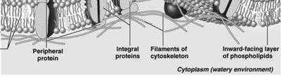
Phospholipid Bilayer: Forms the fundamental structure, with hydrophilic (water-loving) heads facing outward and hydrophobic (water-fearing) tails facing inward.
Membrane Proteins: Integral and peripheral proteins are interspersed throughout the bilayer, contributing to its mosaic appearance and diverse functions.
Cholesterol: Stabilizes membrane fluidity and structure (about 20% of membrane lipids).
Phospholipids: Structure and Importance
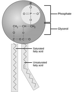
Phospholipids are amphipathic molecules, meaning they have both hydrophilic and hydrophobic regions. This property is essential for forming the selectively permeable barrier of the cell membrane.
Hydrophilic Head: Attracted to water; faces the aqueous environments inside and outside the cell.
Hydrophobic Tails: Repelled by water; face inward, away from the aqueous environments.
Self-Assembly: In aqueous solutions, phospholipids spontaneously form bilayers, creating a stable boundary for cells.
Selective Permeability
The phospholipid bilayer is selectively permeable, allowing only certain molecules to pass directly through.
Can Pass: Small, nonpolar molecules (e.g., O2, CO2).
Cannot Pass: Polar molecules, charged particles (e.g., Na+, K+), and large molecules.
This selective permeability enables cells to control their internal environment and respond to changes outside the cell.
Membrane Proteins: Types and Functions
Membrane proteins are crucial for the diverse functions of the cell membrane. They are classified based on their association with the lipid bilayer:
Integral Proteins: Firmly embedded within the membrane; often span the entire bilayer (transmembrane proteins).
Peripheral Proteins: Loosely attached to the membrane surface or to integral proteins.
Functions of Membrane Proteins
Transport: Channels and carriers move substances across the membrane.
Enzymatic Activity: Catalyze chemical reactions at the membrane surface.
Signal Transduction: Relay signals from the external environment to the cell's interior.
Cell-Cell Recognition: Glycoproteins serve as identification tags for cell recognition.
Intercellular Joining: Form junctions between adjacent cells.
Attachment to Cytoskeleton and Extracellular Matrix: Maintain cell shape and stabilize membrane location.

Enzymes in the Membrane
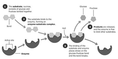
Enzymes are proteins that act as biological catalysts, increasing the rate of chemical reactions without being consumed. Many enzymes are membrane-bound, facilitating reactions at the cell surface.
Active Site: The region of the enzyme where substrates bind and reactions occur.
Enzyme-Substrate Complex: Temporary association between enzyme and substrate during catalysis.
Product Release: Enzyme releases the product and is free to catalyze another reaction.
Cellular Junctions (Cell-Cell Junctions)
Overview of Cell Junctions
Cell junctions are specialized structures that connect adjacent cells, providing structural integrity and facilitating communication. There are three main types:
Tight Junctions
Desmosomes
Gap Junctions
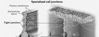
Tight Junctions
Tight junctions are formed by interlocking junctional proteins that seal the space between adjacent cells. They create impermeable barriers that prevent the passage of water and water-soluble substances between cells.
Function: Maintain distinct compartments within tissues; critical in the brain (blood-brain barrier), intestines, and kidneys.
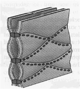
Desmosomes
Desmosomes are anchoring junctions that mechanically attach adjacent cells, providing resistance to mechanical stress. They are composed of plaques, linker proteins, and intermediate filaments.
Function: Abundant in tissues subjected to stretching and mechanical stress, such as skin and heart muscle.
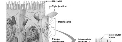
Gap Junctions
Gap junctions are formed by proteins that create channels (connexons) between adjacent cells, allowing direct passage of ions and small molecules. They are essential for cell-to-cell communication and coordination.
Function: Enable synchronized electrical activity, especially important in cardiac muscle tissue.
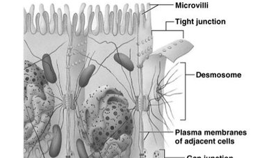
Summary Table: Types of Cell Junctions
Junction Type | Main Structure | Function | Example Location |
|---|---|---|---|
Tight Junction | Interlocking proteins | Seals space between cells; prevents leakage | Intestines, brain, kidneys |
Desmosome | Plaques, linker proteins, intermediate filaments | Anchors cells together; resists mechanical stress | Skin, heart muscle |
Gap Junction | Connexon channels | Allows passage of ions/small molecules; cell communication | Heart, smooth muscle |
Additional info: Cell junctions are critical for tissue integrity and function. Disruption of these junctions can lead to diseases such as blistering disorders (desmosome defects) or impaired organ function (tight junction defects).
