 Indietro
Chapter 3: Cells – The Living Units (Anatomy & Physiology Study Notes)
Guida di studio - Note intelligenti
 Appunti personalizzati basati sui tuoi materiali, ampliati con definizioni chiave, esempi e contesto.
Cells: The Living Units
Definition and Characteristics of a Cell
Cells are the fundamental structural and functional units of life. The human body contains over 250 different types of cells, each specialized in size, shape, and subcellular components to perform unique functions. The cell theory states that the activity of an organism depends on the collective activities of its cells.
Generalized Cell Structure: All human cells share three basic parts: the plasma membrane (outer boundary), cytoplasm (intracellular fluid with organelles), and nucleus (DNA-containing control center).
Cell Diversity: Cells are specialized for various functions such as connecting body parts, movement, storage, defense, information gathering, and reproduction.
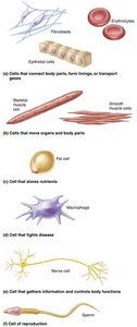
Generalized Cell Structure
The generalized cell contains several key components, each with specific functions essential for cellular life and homeostasis.
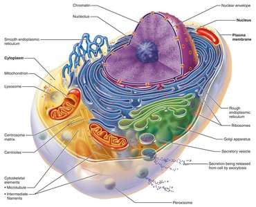
Plasma Membrane: Structure and Function
Overview of the Plasma Membrane
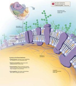
The plasma membrane, also known as the cell membrane, is a dynamic barrier that separates the intracellular fluid (ICF) from the extracellular fluid (ECF). It is primarily composed of a phospholipid bilayer with embedded proteins and cholesterol, and is involved in regulating the entry and exit of substances, cell communication, and cell adhesion.
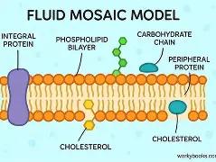
Phospholipid Bilayer
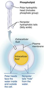
The plasma membrane's basic structure is a bilayer of phospholipids. Each phospholipid molecule has a polar, hydrophilic head and two nonpolar, hydrophobic fatty acid tails. This arrangement creates a semi-permeable barrier between the cell's interior and exterior environments.
Hydrophilic heads: Face outward toward water-containing environments.
Hydrophobic tails: Face inward, away from water.
Membrane Proteins
Membrane proteins are crucial for cell communication, transport, and structural support. They are classified as:
Integral proteins: Span the membrane and function as transporters, enzymes, or receptors.
Peripheral proteins: Loosely attached to the membrane, functioning as enzymes, motor proteins, or in cell-to-cell connections.
Specialized Membrane Structures
Glycocalyx: A carbohydrate-rich area on the cell surface, serving as a biological marker for cell recognition and immune response.
Cell Junctions: Structures such as tight junctions, desmosomes, and gap junctions that connect adjacent cells and facilitate communication or structural integrity.
Transport Across the Plasma Membrane
Passive Transport
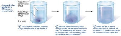
Passive transport involves the movement of substances across the membrane without energy input. It relies on diffusion, where molecules move from areas of high concentration to low concentration (down their concentration gradient).
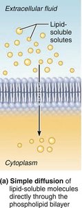
Simple Diffusion: Lipid-soluble molecules and small molecules pass directly through the lipid bilayer.
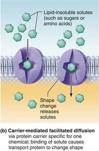
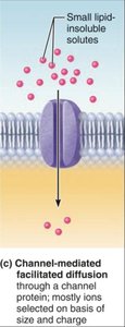
Facilitated Diffusion: Water-soluble or larger molecules cross with the help of carrier or channel proteins.
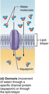
Osmosis: The diffusion of water across a selectively permeable membrane.
Osmosis and Osmotic Pressure
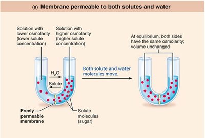
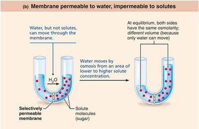
Osmosis is vital for maintaining fluid balance in cells. Water moves from areas of low solute concentration (high water) to high solute concentration (low water) until equilibrium is reached. Osmotic pressure is the force driving water into the cell, while hydrostatic pressure opposes it.
Tonicity and Its Effects on Cells
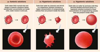
Tonicity describes how a solution affects cell volume:
Isotonic: No net water movement; cell volume remains unchanged.
Hypertonic: Water leaves the cell; cell shrinks (crenation).
Hypotonic: Water enters the cell; cell swells and may burst (lysis).
Active Transport
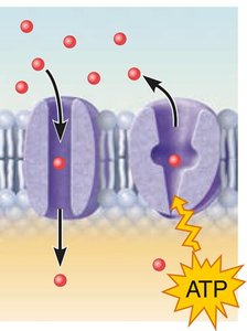
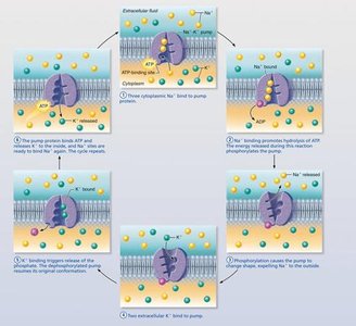
Active transport requires energy (ATP) to move substances against their concentration gradients. It is essential for maintaining cellular homeostasis, especially in excitable cells like nerves and muscles.
Primary Active Transport: Direct use of ATP to transport molecules (e.g., sodium-potassium pump).
Secondary Active Transport: Uses energy stored in ion gradients created by primary active transport.
Vesicular Transport
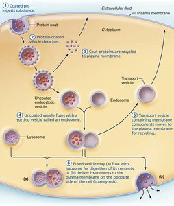
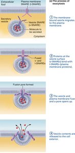
Vesicular transport moves large particles or volumes across membranes using vesicles. Types include:
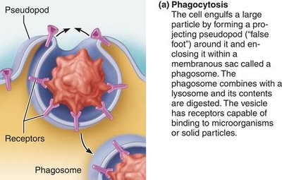
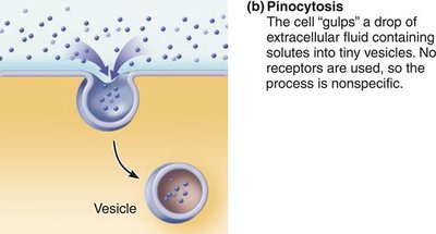
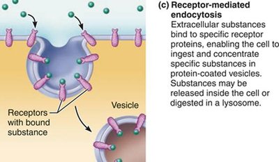
Endocytosis: Bringing substances into the cell (phagocytosis, pinocytosis, receptor-mediated endocytosis).
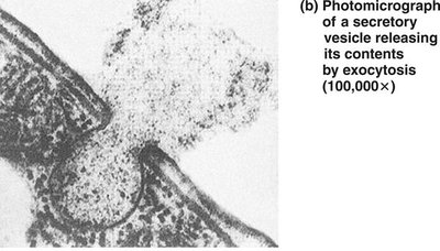
Exocytosis: Expelling substances from the cell.
Transcytosis: Moving substances into, across, and out of the cell.
Cytoplasm and Organelles
Cytoplasm
The cytoplasm is the cellular material between the plasma membrane and the nucleus. It consists of cytosol (fluid), inclusions (stored nutrients or pigments), and organelles (specialized structures).
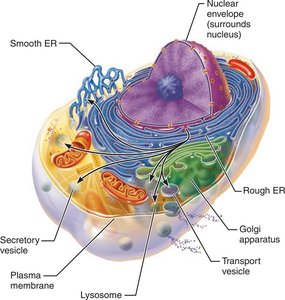
Membranous Organelles
Mitochondria: Site of ATP production via aerobic respiration.
Endoplasmic Reticulum (ER): Rough ER synthesizes proteins; Smooth ER synthesizes lipids and detoxifies chemicals.
Golgi Apparatus: Modifies, sorts, and packages proteins and lipids.
Lysosomes: Digest cellular debris and foreign substances.
Peroxisomes: Neutralize free radicals and detoxify harmful substances.
Non-Membranous Organelles
Ribosomes: Sites of protein synthesis.
Cytoskeleton: Provides structural support and facilitates movement.
Centrioles: Organize spindle fibers during cell division.
Cellular Extensions
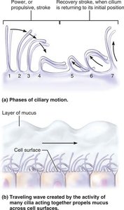
Cilia: Motile extensions that move substances across cell surfaces.
Flagella: Longer extensions that propel cells (e.g., sperm).
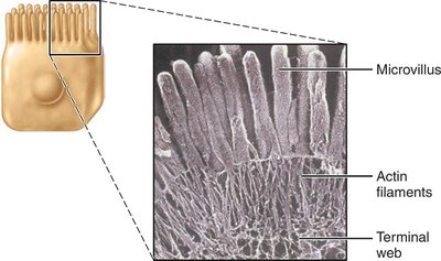
Microvilli: Increase surface area for absorption.
Nucleus: Organization and Function
Structure of the Nucleus
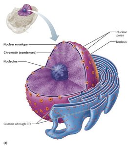
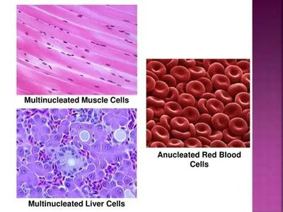
The nucleus is the largest organelle, containing the genetic material (DNA) necessary for protein synthesis and cell function. Most cells are uninucleate, but some are multinucleate or anucleate.
Nuclear Envelope: Double membrane surrounding the nucleus.
Nucleoli: Sites of ribosomal RNA synthesis.
Chromatin: DNA and protein complex; condenses to form chromosomes during cell division.
Cell Cycle and Division
The cell cycle consists of interphase (growth and normal function) and the mitotic phase (cell division). Mitosis divides the nucleus, while cytokinesis divides the cytoplasm. Meiosis is a special division for gametes.
Genetic Material and Protein Synthesis
DNA contains the genetic code for protein synthesis. Genes are segments of DNA coding for polypeptides. RNA acts as the intermediary, transcribing and translating the genetic code into proteins.
Transcription: DNA code is copied into mRNA in the nucleus.
Translation: mRNA is decoded at ribosomes to assemble amino acids into proteins.
Cell Metabolism and Homeostasis
Clinical Implications
Hyperplasia: Increased cell production, as seen in anemia, to restore homeostasis.
Atrophy: Decreased cell size or number due to reduced stimulation (e.g., muscular dystrophy).
Progeria: Rare disease causing premature aging due to defective nuclear proteins.
Summary Table: Types of Membrane Transport
Transport Type | Energy Required? | Direction | Examples |
|---|---|---|---|
Simple Diffusion | No | High to Low | O2, CO2, steroids |
Facilitated Diffusion | No | High to Low | Glucose, amino acids |
Osmosis | No | High to Low (water) | Water |
Active Transport | Yes (ATP) | Low to High | Na+/K+ pump |
Vesicular Transport | Yes (ATP) | Varies | Endocytosis, exocytosis |
Additional info: This guide expands on the provided lecture content with definitions, examples, and clinical implications to ensure a comprehensive understanding of cell structure and function for Anatomy & Physiology students.
