 Indietro
Chapter 4: Tissues – The Living Fabric (Anatomy & Physiology Study Notes)
Guida di studio - Note intelligenti
 Appunti personalizzati basati sui tuoi materiali, ampliati con definizioni chiave, esempi e contesto.
Principles of Structure and Function
Levels of Organization
The human body is organized into hierarchical levels, each increasing in complexity:
Chemical Level: Atoms and molecules form the basis of life.
Cellular Level: Cells are the basic structural and functional units.
Tissue Level: Groups of similar cells working together for a specific function.
Organ Level: Structures composed of multiple tissue types performing specific tasks.
Organ System Level: Groups of organs working together for major body functions.
Organism Level: The complete living individual.
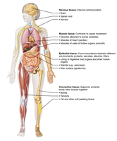
Overview of Tissue Types
Tissues are groups of similar cells that perform a common function. The study of tissues is called histology.
Epithelial Tissue: Covers surfaces, lines cavities, forms glands.
Connective Tissue: Supports, protects, binds other tissues.
Muscle Tissue: Contracts to produce movement.
Nervous Tissue: Internal communication via electrical impulses.
Epithelial Tissue
General Characteristics
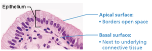
Epithelial tissues form continuous sheets and serve as coverings/linings or as glandular structures. Their functions include protection, absorption, secretion, filtration, excretion, and sensory reception. The specific function depends on the type of epithelium.
Polarity: Distinct apical (exposed) and basal (attached) surfaces.
Basement Membrane: Composed of basal lamina (adhesive) and reticular lamina (collagen fibers).
Tightly Packed Cells: Connected by tight junctions and desmosomes.
Avascular: No blood vessels; nutrients diffuse from underlying tissues.
Innervated: Contains nerve fibers.
Regenerative: Rapid cell division for repair.
Classification of Epithelial Tissue
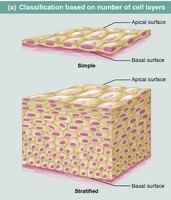
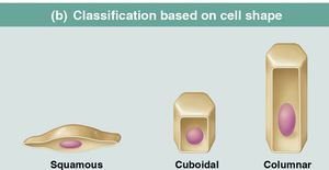
Epithelial tissues are classified by the number of cell layers and the shape of cells:
Number of Layers:
Simple: One layer of cells.
Stratified: Multiple layers of cells.
Cell Shape:
Squamous: Flat, scale-like.
Cuboidal: Cube-shaped.
Columnar: Tall, column-like.
Types of Epithelial Tissue
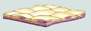
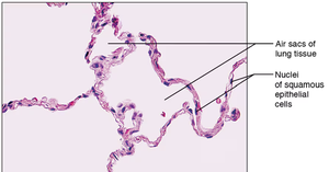
Simple Squamous Epithelium
Single layer of flat cells. Found in blood vessels, alveoli, glomeruli, and serosae. Functions include filtration, diffusion, and secretion.
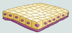
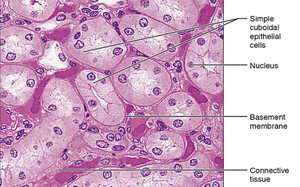
Simple Cuboidal Epithelium
Single layer of cube-shaped cells. Located in kidney tubules, surfaces of ovaries. Functions: secretion and absorption.
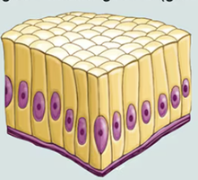
Simple Columnar Epithelium
Single layer of tall, column-like cells. Found in most of the digestive tract and uterine tubes. Functions: absorption, secretion, movement of mucus. May have cilia or microvilli.
Connective Tissue
General Characteristics
Connective tissues are the most abundant and widely distributed tissues in the body. They support, protect, insulate, store energy, and transport substances. All connective tissues share:
Extracellular Matrix (ECM): Non-living material outside cells, composed of ground substance and fibers.
Cells: Produce ECM (except blood cells).
Common Origin: Derived from embryonic mesenchyme.
Vascularity: Varies by tissue type.
Structural Components
Ground Substance: Contains interstitial fluid, cell adhesion proteins, proteoglycans/glycosaminoglycans (increase viscosity).
Fibers:
Collagen: Strong, thick, most numerous.
Elastic: Thin, stretch and recoil.
Reticular: Thin collagen, form nets.
Cells:
Fibroblasts/Fibrocytes: In connective tissue proper.
Chondroblasts/Chondrocytes: In cartilage.
Osteoblasts/Osteocytes: In bone.
Adipocytes: Fat storage.
White Blood Cells, Mast Cells, Macrophages: Immune functions.
Muscle Tissue
Types and Characteristics
Muscle tissue is specialized for contraction and movement. It contains tightly packed cells with myofilaments (actin and myosin).
Skeletal Muscle: Voluntary, striated, multi-nucleated, attached to bones.
Cardiac Muscle: Involuntary, striated, branched cells with intercalated discs, found in heart wall.
Smooth Muscle: Involuntary, non-striated, tapered cells, found in walls of hollow organs and blood vessels.
Nervous Tissue
Cell Types and Functions
Nervous tissue is responsible for internal communication. It consists of:
Neurons: Transmit electrical impulses, sensory reception, integration, control of effectors.
Neuroglia (Glial Cells): Support, protect, and clean up.
Locations include the brain, spinal cord, and nerves.
Membranes
Epithelial Tissue Membranes
Cutaneous Membrane: Skin; stratified squamous epithelium (epidermis) and dense irregular connective tissue (dermis).
Mucous Membranes: Line cavities open to exterior; stratified squamous or simple columnar epithelium and areolar connective tissue (lamina propria).
Serous Membranes: Line enclosed cavities; simple squamous epithelium (mesothelium) and areolar connective tissue. Double membrane structure (visceral and parietal layers).
Connective Tissue Membrane
Synovial Membrane: Lines joint cavities; composed solely of connective tissue.
Tissue Damage & Repair
Steps of Tissue Repair
Inflammation: Release of histamine and heparin, increased capillary permeability, WBCs and clotting factors enter area, edema, blood clotting, scab formation.
Organization: Restoration of blood supply, clot replaced by granulation tissue, fibroblasts produce collagen, macrophages clean debris, epithelial cells cover granulation tissue.
Regeneration and Fibrosis: Epithelium regenerates, scar tissue forms, wound margins contract, scab detaches.
Review Questions
An epithelium that has several layers, with an apical layer of flattened cells, is called:
Stratified Squamous Epithelium
Compare and contrast skeletal, cardiac, and smooth muscle tissue relative to structure, body location, and specific function.
Skeletal: Striated, voluntary, multi-nucleated, attached to bones, movement and posture.
Cardiac: Striated, involuntary, branched, single nucleus, heart wall, pumps blood.
Smooth: Non-striated, involuntary, tapered, single nucleus, walls of organs/vessels, moves substances.
The gland type that secretes products such as milk, saliva, bile, or sweat through a duct is:
Exocrine gland
Polarity is a property of all normal epithelial tissues.
True
A sebaceous (oil) gland of the skin must completely rupture to release its secretory products. Thus, this type of gland is considered to be:
Holocrine gland
The skin is a _____________ membrane.
Cutaneous
Additional info: Academic context and explanations have been expanded for clarity and completeness. Only images directly relevant to the explanation have been included.
