 Indietro
Chapter 6: The Integumentary System – Structure, Function, and Clinical Relevance
Guida di studio - Note intelligenti
 Appunti personalizzati basati sui tuoi materiali, ampliati con definizioni chiave, esempi e contesto.
The Integumentary System
Overview and Organization
The integumentary system consists of the skin and its accessory structures, including hair, nails, glands, and sensory receptors. It is the largest organ system in the body and serves as a protective barrier, regulates temperature, and facilitates sensory perception.
Tissue: Group of cells performing a similar function.
Organ: Two or more types of tissues grouped together for specialized functions.
Organ System: Composed of various organs and structures performing a specific body process.
Skin: Composed of epithelial tissue (epidermis) overlying connective tissue (dermis).
Cutaneous membrane: Another term for skin.
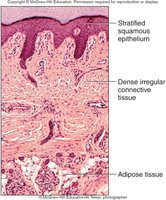
Layers of the Integument
The skin is organized into three main layers: the epidermis, dermis, and subcutaneous layer (hypodermis). Each layer has distinct structural and functional properties.
Epidermis: Stratified squamous epithelium; outermost layer.
Dermis: Dense irregular connective tissue; deeper layer.
Subcutaneous layer (hypodermis): Areolar and adipose connective tissue; not part of the integumentary system but provides insulation and energy storage.
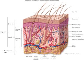
Functions of the Integument
Protective and Regulatory Functions
The integument serves multiple essential functions for the body, including protection, regulation, and synthesis.
Protection: Shields the body from injury, harmful substances, microbes, extreme temperatures, and UV radiation.
Prevention of water loss/gain: Epidermis is water-resistant; water is lost via sweat and transpiration.
Vitamin D synthesis: Formation of vitamin D3 (cholecalciferol), a precursor to calcitriol, which regulates calcium and phosphate absorption.
Secretion: Waste products are secreted onto the skin surface during sweating.
Absorption: Skin absorbs some chemicals/drugs; suitable for transdermal administration.
Immune function: Dendritic cells initiate immune responses.
Temperature regulation: Dermal blood vessels vasoconstrict or vasodilate to conserve or release heat.
Sensory reception: Receptors detect stimuli such as touch and pressure.
Structure of the Epidermis
Layers (Strata) of the Epidermis
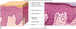
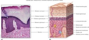
The epidermis is a keratinized, stratified squamous epithelium composed of several distinct layers, each with specialized functions.
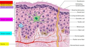
Stratum basale: Deepest layer; single layer of cuboidal cells; only mitotic layer.
Stratum spinosum: Several layers of polygonal keratinocytes; new cells from stratum basale.
Stratum granulosum: 3–5 layers; keratinocytes begin keratinization; cells die.
Stratum lucidum: Translucent, 2–3 cell layers thick; found only in thick skin (palms, soles).
Stratum corneum: Outermost layer; 20–30 layers of dead, keratinized cells; protective.
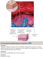
Keratinized Stratified Squamous Epithelium
Keratinized stratified squamous epithelium is characterized by multiple cell layers, with superficial cells lacking nuclei and filled with keratin. This structure provides protection against abrasion and infection.
Keratinocytes: Found in all layers; synthesize keratin; migrate toward the surface and die.
Function: Protection of underlying tissues from abrasion.
Specialized Cells of the Epidermis
The epidermis contains several specialized cell types, each with unique functions.
Dendritic (Langerhans) cells: Found in stratum spinosum; phagocytes; protect against infection.
Tactile (Merkel) cells: Found in stratum basale; sensory receptors for light touch.
Melanocytes: Found in stratum basale; produce melanin, which absorbs UV light and provides skin color.
Skin Color and Clinical Relevance
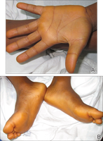
Skin color is determined by three main pigments: hemoglobin, melanin, and carotene. Variations in these pigments can have clinical significance.
Hemoglobin: Oxygen-binding protein; gives skin a reddish hue.
Melanin: Produced by melanocytes; provides brown, black, tan, yellow, or red shades.
Carotene: Yellow-orange pigment from diet; excessive intake can cause carotenemia.
Albinism: Melanocytes unable to produce melanin.
Structure of the Dermis
Layers of the Dermis
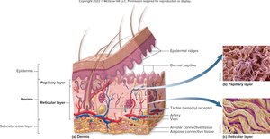
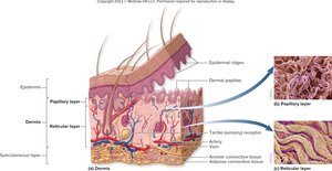
The dermis is deep to the epidermis and consists of two layers: the papillary layer and the reticular layer. It contains blood vessels, glands, hair follicles, and sensory nerve endings.
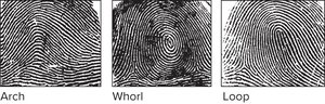
Papillary layer: Superficial; areolar connective tissue; contains dermal papillae that interlock with epidermal ridges, forming fingerprints.
Reticular layer: Deeper; dense irregular connective tissue; provides strength and elasticity.
Friction Ridges and Lines of Cleavage
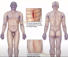
Friction ridges are patterns formed by dermal papillae, visible as fingerprints. Lines of cleavage (Langer's lines) are patterns of collagen and elastic fibers that influence wound healing and surgical incisions.
Friction ridges: Enhance grip and tactile sensation.
Lines of cleavage: Incisions parallel to these lines heal better; perpendicular incisions may gape.
Stretch marks (striae): Result from overstretching and tearing of collagen fibers.
Subcutaneous Layer (Hypodermis)
Structure and Function
The subcutaneous layer lies deep to the dermis and is composed of areolar and adipose connective tissue. It is not part of the integumentary system but plays important roles in protection, energy storage, and insulation.
Functions: Protection, energy storage, insulation, and rapid absorption of injected drugs.
Thickness/distribution: Influenced by sex hormones.
Accessory Structures of the Skin
Hair
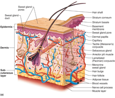
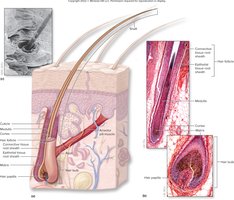
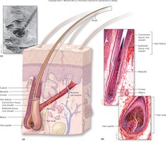
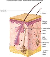
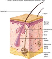
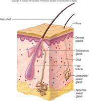
Hair is found almost everywhere on the body and is composed of keratinized cells. It grows from hair follicles and serves various functions.
Types of hair: Lanugo (fetal), vellus (fine, covers limbs), terminal (coarse, pigmented, scalp, eyebrows, eyelashes).
Zones of hair: Hair bulb (living cells), root (from bulb to skin surface), shaft (beyond skin surface, dead cells).
Functions: Protection, heat retention, sensory reception, visual identification.

Hair Growth and Clinical Conditions
Hair growth and loss are influenced by genetic, hormonal, and environmental factors. Several clinical conditions affect hair.
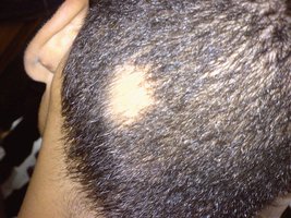
Alopecia areata: Autoimmune disorder causing patchy hair loss.
Diffuse hair loss: Hair shed from all parts of scalp; due to hormones, drugs, iron deficiency.
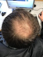
Androgenic alopecia: Male pattern baldness; DHT causes miniaturization of hair follicles.
Exocrine Glands of the Skin
The skin contains several types of exocrine glands, each with distinct functions and secretions.
Sweat glands: Merocrine (eccrine) and apocrine types; aid in thermoregulation and waste excretion.
Merocrine glands: Most numerous; secrete watery sweat directly onto skin surface.
Apocrine glands: Secrete viscous, cloudy sweat into hair follicles; produce odor when acted on by bacteria.
Sebaceous glands: Holocrine glands; secrete oily sebum into hair follicles; lubricates skin and hair.
Ceruminous glands: Modified apocrine glands; produce earwax (cerumen).
Mammary glands: Modified apocrine glands; produce milk in lactating females.
Chemical Reactions and Enzymes in the Skin
Types of Chemical Reactions
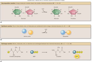
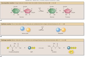
Chemical reactions in the body include decomposition, synthesis, and exchange reactions. Enzymes catalyze these reactions, lowering activation energy and increasing reaction rates.
Decomposition reaction: AB → A + B; e.g., hydrolysis of sucrose.
Synthesis reaction: A + B → AB; e.g., dehydration synthesis forming a dipeptide.
Exchange reaction: Atoms exchanged between molecules; e.g., creatine phosphate and ADP.
Enzyme Function and Structure
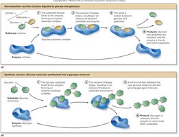
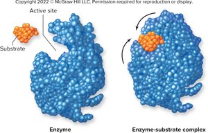
Enzymes are proteins that act as biological catalysts. They are highly specific, with active sites that bind only certain substrates.
Decrease activation energy: Facilitate reactions that would occur naturally.
Increase rate of product formation: Essential for cellular metabolism.
Naming: Based on substrate or product; suffix -ase (e.g., lactase, DNA polymerase).
Clinical View: Lactose Intolerance
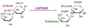
Lactose intolerance is caused by a deficiency or abnormality in lactase, the enzyme required to break down lactose into glucose and galactose. Symptoms include abdominal upset, nausea, diarrhea, bloating, and gas.
Treatment: Lactase supplements, avoidance of milk, or lactose-free milk.

Vitamin D Production and Clinical Relevance
Production of Vitamin D
Vitamin D is synthesized in the skin upon exposure to UV light, then converted to its active forms in the liver and kidneys. Calcitriol regulates calcium absorption.
Step 1: UV light converts 7-dehydrocholesterol to vitamin D3 (cholecalciferol).
Step 2: Vitamin D3 converted to calcidiol in the liver.
Step 3: Calcidiol converted to calcitriol in the kidneys.
Function: Calcitriol controls calcium absorption.
Vitamin D Deficiency
Low levels of vitamin D can lead to various skin conditions and are influenced by age, diet, and reduced enzyme activity in the liver and kidneys.
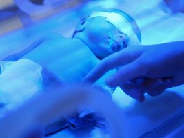
Associated conditions: Psoriasis, atopic dermatitis, acne vulgaris, vitiligo, jaundice.
Causes: Reduced skin production, decreased intestinal absorption, decreased enzyme activity.
Repair and Regeneration of the Integumentary System
Tissue Repair Mechanisms
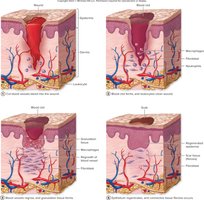
Tissue repair occurs via regeneration or fibrosis. Regeneration restores function by replacing damaged cells with the same type, while fibrosis fills gaps with scar tissue, which does not restore function.
Regeneration: Replacement of damaged cells; restores function.
Fibrosis: Scar tissue formation; functional activities not restored.
Healing of Wounds and Burns
Wound healing involves inflammation, clot formation, re-growth of blood vessels, and regeneration of epithelium and connective tissue. Burns are classified by depth and severity.
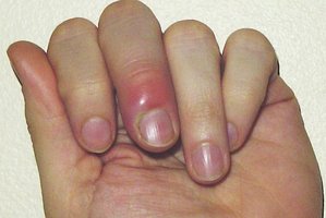
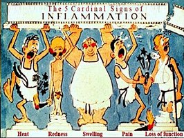
Inflammation: Body's response to injury; restricts spread of infection.
Stages of wound healing: Bleeding, clot formation, granulation tissue, regeneration, fibrosis.
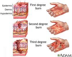
Burns: First degree (epidermis), second degree (epidermis and dermis), third degree (epidermis, dermis, subcutaneous layer).
Rule of nines: Used to estimate surface area of burns.
Layer | Structure | Function |
|---|---|---|
Epidermis | Stratified squamous epithelium | Protection, water resistance |
Dermis | Dense irregular connective tissue | Strength, elasticity, houses glands and follicles |
Hypodermis | Adipose and areolar tissue | Insulation, energy storage |
Burn Degree | Layers Involved | Symptoms |
|---|---|---|
First | Epidermis | Redness, pain |
Second | Epidermis, part of dermis | Blistering, pain, slight scarring |
Third | Epidermis, dermis, hypodermis | Severe scarring, dehydration, infection risk |
Additional info: Academic context was added to clarify the structure and function of skin layers, accessory structures, and clinical relevance of skin conditions.
