 Indietro
Chapter 8: The Nervous System – Anatomy & Physiology Study Guide
Guida di studio - Note intelligenti
 Appunti personalizzati basati sui tuoi materiali, ampliati con definizioni chiave, esempi e contesto.
The Nervous System: Overview
Organ Systems That Coordinate
The nervous system and endocrine system are responsible for maintaining homeostasis by coordinating activities throughout the body. The nervous system is the most complex organ system, providing rapid, brief, and specific responses, while the endocrine system produces slower, longer-lasting, and more general effects.
Nervous System: Fast, brief, specific responses.
Endocrine System: Slow, long-lasting, general effects.
Nervous System Functions
The nervous system performs three main functions:
Monitoring: Internal and external environments.
Integration: Sensory information.
Coordination: Voluntary and involuntary responses.
Anatomical and Functional Divisions of the Nervous System
Central Nervous System (CNS) and Peripheral Nervous System (PNS)
The nervous system is divided into the CNS and PNS. The CNS includes the brain and spinal cord, responsible for integrating and coordinating sensory processing and motor transmission. The PNS consists of all neural tissues outside the CNS, connecting the CNS to the rest of the body.
CNS: Brain and spinal cord; higher functions (intelligence, memory, emotion).
PNS: Neural tissues outside CNS; communication between CNS and body.
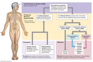
PNS Functional Divisions
Afferent Division: Sensory; brings information to CNS from receptors.
Efferent Division: Motor; carries information from CNS to effectors (muscles and glands).
Efferent Motor Subdivisions
Somatic Nervous System (SNS): Controls skeletal muscle.
Autonomic Nervous System (ANS): Controls smooth and cardiac muscle, glands; includes sympathetic and parasympathetic divisions.
Neural Tissue: Structure and Function
Types of Neural Cells
Neurons: Basic functional units; communicate with other cells.
Neuroglia (Glial Cells): Support and regulate environment around neurons; more numerous and capable of division.
General Structure of Neurons
Neurons consist of a cell body, dendrites (receive signals), axon (carries signals away), and axon terminals (form synapses).
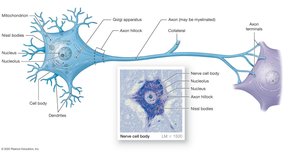
Cell Body: Contains nucleus, nucleolus, mitochondria, ribosomes, rough ER (Nissl bodies).
Dendrites: Receive incoming signals.
Axon: Transmits signals away from cell body.
Axon Hillock: Origin of action potentials.
Neuron Regeneration
Most CNS neurons lack centrioles and cannot divide.
Neural stem cells are typically inactive unless injury occurs.
Structural Classification of Neurons
Multipolar: Two or more dendrites, one axon; motor neurons and CNS.
Unipolar: Dendrites and axon continuous; most sensory neurons.
Bipolar: One dendrite, one axon; special sense organs.
Functional Classification of Neurons
Sensory (Afferent) Neurons: Receive information from receptors, relay to CNS.
Motor (Efferent) Neurons: Carry instructions from CNS to effectors.
Interneurons: Located in CNS; interconnect neurons, involved in higher functions.
Neuroglial Cells
CNS: Astrocytes, oligodendrocytes, microglia, ependymal cells.
PNS: Satellite cells, Schwann cells.
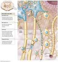
Astrocytes (CNS)
Star-shaped, largest and most numerous.
Maintain blood-brain barrier, isolate CNS from circulation.
Oligodendrocytes (CNS)
Produce myelin, insulating axons.
Myelinated areas = white matter; unmyelinated = gray matter.
Microglia (CNS)
Smallest, least numerous; phagocytic, engulf pathogens and waste.
Ependymal Cells (CNS)
Line cavities filled with cerebrospinal fluid (CSF); involved in CSF production and circulation.
Schwann Cells (PNS)
Cover every axon in PNS; form myelin sheath or neurilemma.
One Schwann cell can encircle several unmyelinated axons.
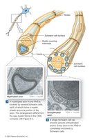
Satellite Cells (PNS)
Surround and support neuron cell bodies; similar to astrocytes.
Organization of Neurons
PNS Organization
Ganglia: Collections of neuron cell bodies.
Nerves: Bundles of axons; spinal nerves (spinal cord), cranial nerves (brain).
CNS Organization
Centers/Nuclei: Collections of cell bodies (gray matter).
Neural Cortex: Thick layer of gray matter on brain surface.
Tracts: Bundles of axons (white matter); columns in spinal cord.
Pathways: Sensory (ascending) and motor (descending).
Membrane Potential and Action Potentials
Membrane Potential of a Neuron
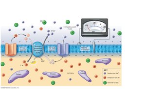
Neurons have a polarized plasma membrane, with positive charges outside and negative charges inside. The potential difference is called membrane potential, measured in millivolts. Resting membrane potential is −70 mV.
Factors Influencing Membrane Potential
Imbalance in electrical charges.
Extracellular fluid: high in Na+ and Cl−.
Intracellular fluid: high in K+ and negatively charged proteins.
Ions move via leak channels (always open) and gated channels (open/close under specific conditions).
Passive Ion Movement
Sodium moves into cell (chemical and electrical gradient).
Potassium moves out (chemical gradient stronger than electrical).
Potassium diffuses out faster than sodium enters.
Maintaining Resting Membrane Potential
Active transport via sodium-potassium exchange pump.
Pumps 3 Na+ out for every 2 K+ in.
Net loss of positive ions results in −70 mV resting potential.
Changes in Membrane Potential
Stimuli alter permeability to Na+ or K+, or activity of exchange pump.
Opening Na+ channels causes depolarization (toward 0 mV).
Opening K+ channels causes repolarization or hyperpolarization (more negative).
Graded Potentials
Local changes in membrane potential; decrease with distance from stimulus.
Can trigger action potentials in large neurons/muscle fibers.
Action Potentials
Action potentials are propagated changes in membrane potential of excitable cells, traveling the entire length of the cell. They follow the all-or-none principle and are initiated when threshold is reached.
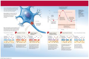
Threshold: Level of depolarization required to initiate action potential.
Refractory Period: Time during which membrane cannot respond to further stimulation.
Generation and Propagation of Action Potentials
Begins at axon hillock; depolarizes to threshold (−60 mV).
Voltage-gated Na+ channels open, depolarize to +30 mV.
Na+ channels close, K+ channels open, repolarize membrane.
K+ channels close, membrane returns to resting potential.
Propagation Types
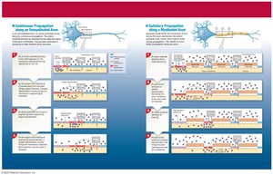
Continuous Propagation: Unmyelinated fibers; slow (1 m/s).
Saltatory Propagation: Myelinated axons; fast (18–140 m/s); depolarization occurs only at nodes of Ranvier.
Synapses and Neurotransmitters
The Synapse
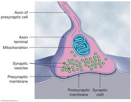
A synapse is the site where a neuron communicates with another cell via neurotransmitters. Communication is unidirectional.
Presynaptic Neuron: Sends information; axon terminal contains neurotransmitter vesicles.
Postsynaptic Neuron: Receives information; has receptors for neurotransmitters.
Acetylcholine (ACh)
Activates cholinergic synapses in four steps: arrival of action potential, release and diffusion of ACh, binding to receptors, removal by acetylcholinesterase (AChE).
Other Neurotransmitters
Norepinephrine (NE): Adrenergic synapses; important in brain and ANS.
Dopamine, GABA, Serotonin: CNS neurotransmitters; various effects.
Nitric Oxide (NO), Carbon Monoxide (CO): Gaseous neurotransmitters.
Excitatory and Inhibitory Synapses
ACh and NE usually excitatory (depolarization).
Dopamine, GABA, serotonin usually inhibitory (hyperpolarization).
Effect depends on receptor type.
Postsynaptic Response
Multiple presynaptic neurons can have cumulative effects.
Excitatory inputs can trigger action potential; inhibitory inputs prevent it.
Mixed inputs may cancel each other.
Neuronal Pools
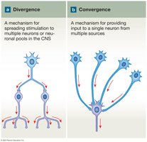
Groups of interconnected interneurons with specific functions.
Divergence: Spreads information from one neuron to multiple neurons.
Convergence: Multiple neurons synapse with a single postsynaptic neuron.
The Brain: Structure and Function
Major Regions of the Brain
Cerebrum
Diencephalon
Midbrain
Pons
Medulla oblongata
Cerebellum
Brain Structure Overview
Cerebrum: Conscious thoughts, sensations, memory, intellect.
Diencephalon: Thalamus, hypothalamus, epithalamus; processing centers, relay station, autonomic functions.
Brain Stem: Midbrain, pons, medulla oblongata; processing centers, relay station, autonomic functions.
Cerebellum: Motor activities, balance.
Ventricles of the Brain
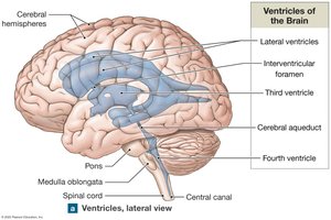
Internal cavities filled with cerebrospinal fluid (CSF), lined with ependymal cells.
Two lateral ventricles, third ventricle (diencephalon), fourth ventricle (between cerebellum and brainstem).
Connected by passageways for CSF flow.
Cerebrospinal Fluid (CSF)
Surrounds and bathes CNS surfaces.
Cushions, supports, transports nutrients, chemicals, and wastes.
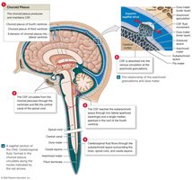
Produced by choroid plexus in each ventricle; 500 mL/day, replaced every 8 hours.
Circulates from fourth ventricle to subarachnoid space and dural sinuses; absorbed by arachnoid granulations.
Cerebrum
Largest brain region; includes gray and white matter.
Surface forms gyri (folds), sulci (shallow depressions), fissures (deep grooves).
Longitudinal fissure divides hemispheres.
Cerebral Hemispheres and Lobes
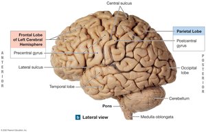
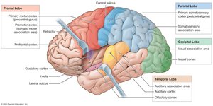
Frontal, parietal, temporal, occipital lobes; insula (deep to temporal lobe).
Each lobe has sensory and motor regions; hemispheres control opposite sides of body.
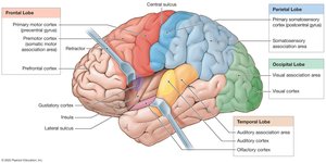
Features of the Cerebral Hemispheres
Central sulcus: divides frontal and parietal lobes.
Lateral sulcus: separates frontal and temporal lobes.
Parieto-occipital sulcus: separates parietal and occipital lobes.
Motor and Sensory Areas
Precentral gyrus (frontal lobe): primary motor cortex (voluntary movement).
Postcentral gyrus (parietal lobe): primary somatosensory cortex (touch, pressure, pain, temperature).
Other Sensory Areas
Visual cortex (occipital lobe): vision.
Gustatory cortex (frontal lobe): taste.
Auditory cortex: hearing.
Olfactory cortex (temporal lobe): smell.
Association Areas
Integrate sensory and motor cortexes; interpret information, coordinate motor response.
Somatosensory association area: recognizes touch.
Premotor cortex: coordinates learned movements.
Cortical Connections
Linked by deeper white matter; hemispheres interconnected via corpus callosum.
Other axons link cortex with diencephalon, brain stem, cerebellum, spinal cord.
Cerebral Processing Centers
Receive information from association areas; direct complex motor activities.
Often lateralized to one hemisphere.
Wernicke’s area: integrates sensory information, memory.
Broca’s area: motor speech, regulates breathing/vocalization.
Prefrontal Cortex
Coordinates information from association areas; abstract intellectual functions, behavior.
Hemispheric Lateralization
Left: language, analytical tasks, logic.
Right: spatial analysis, sensory input, emotional context.
Communication via corpus callosum.
Electroencephalogram (EEG)
Printed record of brain electrical activity; brain waves correlate with consciousness and disorders.
Other mapping: PET, MRI.
Memory
Fact memories: specific information.
Skill memories: learned motor skills.
Short-term: immediate recall; long-term: lasting, consolidated.
Amnesia: memory loss from disease/trauma.
Basal Nuclei
Gray matter beneath lateral ventricles; subconscious control of skeletal muscle tone, coordination of learned movements.
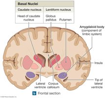
Components of Basal Nuclei
Caudate nucleus, lentiform nucleus, amygdaloid body (limbic system).
Limbic System
Functional grouping; emotional states, links conscious/unconscious, aids memory storage/retrieval (hippocampus).
Diencephalon
Switching/relay centers; integrate sensory/motor commands.
Surrounds third ventricle; epithalamus, thalamus, hypothalamus.
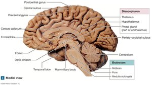
Epithalamus
Anterior: choroid plexus; posterior: pineal gland (melatonin, day-night cycles).
Thalamus
Relay/processing center for sensory information (except olfactory); filters information to cortex.
Hypothalamus
Emotion, autonomic functions, hormone production; pituitary gland link to endocrine system.
Hypothalamus Functions
Subconscious skeletal muscle control (emotion), adjusts pons/medulla, coordinates nervous/endocrine systems, secretes hormones, produces thirst/hunger, regulates temperature, daily cycles.
Midbrain
Nuclei for cranial nerves; reticular formation (wakefulness, involuntary functions); communicates with basal nuclei (dopamine, Parkinson’s disease).
Pons
Links cerebellum with other brain regions; sensory/motor nuclei for cranial nerves; influences respiration.
Cerebellum
Automatic processing; adjusts balance, fine-tunes movements; cerebellar peduncles link cortex, basal nuclei, brain stem; ataxia = balance disturbance.
Medulla Oblongata
Connects brain with spinal cord; sensory/motor nuclei for cranial nerves; reflex centers (cardiac, vasomotor, respiratory rhythmicity).
