 Indietro
Lecture 5 - Histology: The Study of Tissues in Anatomy & Physiology
Guida di studio - Note intelligenti
 Appunti personalizzati basati sui tuoi materiali, ampliati con definizioni chiave, esempi e contesto.
Histology Overview
Definition and Importance
Histology is the study of tissues and their arrangement within organs. Tissues are groups of similar cells and cell products that originate from the same embryonic region and work together to perform specific structural or physiological roles. Understanding tissue structure and function is fundamental to comprehending organ systems in Anatomy & Physiology.
Tissue: A collection of similar cells and cell products performing a specific function.
Four primary tissue types: Epithelial, Connective, Muscular, Nervous.
Tissues differ in cell function, extracellular matrix composition, and the proportion of cells to matrix.
Primary Tissue Classes
Classification and General Functions
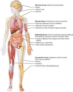
The human body is composed of four primary tissue classes, each with distinct functions and structural characteristics.
Epithelial tissue: Covers surfaces, lines cavities, forms glands.
Connective tissue: Supports, protects, binds other tissues.
Muscular tissue: Contracts to produce movement.
Nervous tissue: Enables internal communication via electrical signals.
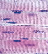
Epithelial Tissue
Structure and Functions
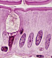
Epithelial tissue forms sheets of cells that cover body surfaces and line cavities. It is essential for protection, secretion, absorption, excretion, filtration, and sensation.
Covering and lining epithelium: Forms skin, lines open cavities, covers organs.
Glandular epithelium: Forms glands for secretion.
Functions: Protection, Secretion, Excretion, Absorption, Filtration, Sensation.
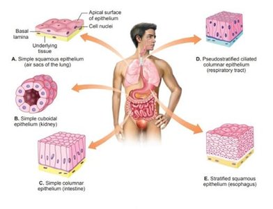
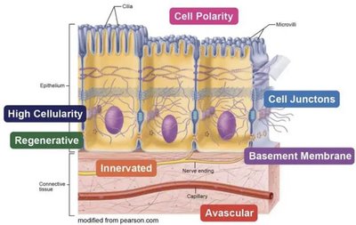
Special Characteristics of Epithelial Tissue
Epithelial tissue exhibits several unique features:
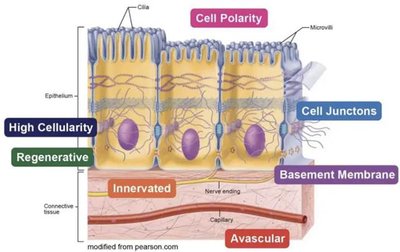
Polarity: Distinct apical (top) and basal (bottom) surfaces.
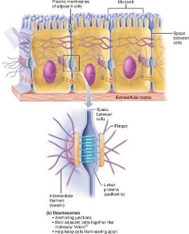
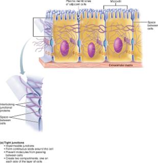
Specialized contacts: Cells are tightly joined by junctions (tight junctions, desmosomes).
Supported by connective tissue: Basement membrane anchors epithelium.
Avascular but innervated: No blood vessels, but has nerve supply.
Regeneration: High capacity for renewal after damage.
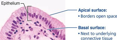
Polarity
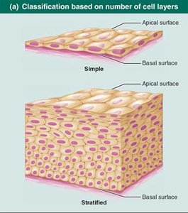
Epithelial cells have an apical surface exposed to a cavity or external environment and a basal surface attached to underlying connective tissue.
Apical surface: Faces open space.
Basal surface: Faces underlying connective tissue.
Specialized Contacts
Epithelial cells are closely packed, forming continuous sheets. Lateral contacts include tight junctions (prevent leakage) and desmosomes (provide mechanical strength).
Tight junctions: Seal cells together.
Desmosomes: Anchor cells to each other.
Support by Connective Tissue
All epithelial sheets rest on a basement membrane, which consists of the basal lamina and reticular lamina (collagen network).
Basement membrane: Provides structural support and regulates exchange.
Avascular but Innervated
Epithelial tissue lacks blood vessels and receives nutrients by diffusion from underlying connective tissue. It is innervated, allowing sensation. 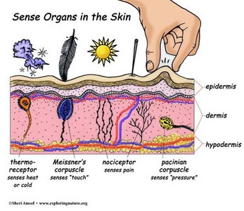
Regeneration
Epithelial cells rapidly regenerate, especially when exposed to friction or hostile environments. 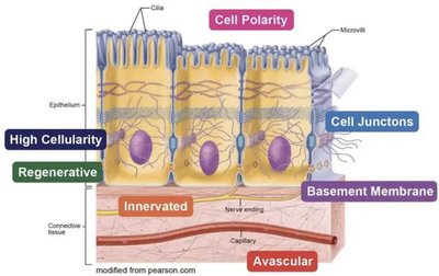
Clinical Homeostatic Imbalances
Cancerous epithelial cells can breach the basement membrane, invading underlying tissues and spreading cancer.
Epithelial Tissue Categories
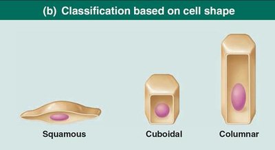
Epithelial tissues are classified by cell layers and cell shape:
Simple epithelium: Single cell layer.
Stratified epithelium: Multiple cell layers.
Squamous: Flattened, scale-like cells.
Cuboidal: Cube-shaped cells.
Columnar: Tall, column-like cells.
Types of Epithelial Tissue
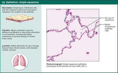
Simple squamous epithelium: Rapid diffusion; found in air sacs of lungs, blood vessels.
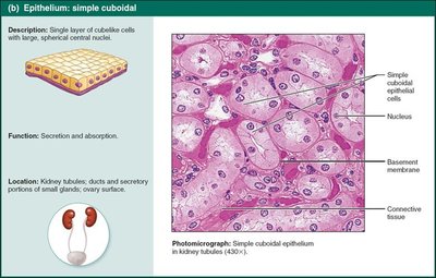
Simple cuboidal epithelium: Secretion and absorption; found in kidney tubules.
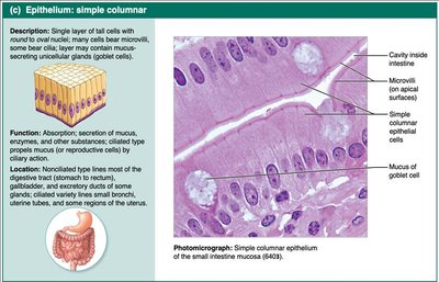
Simple columnar epithelium: Absorption and secretion; found in digestive tract.
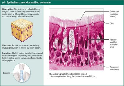
Pseudostratified columnar epithelium: Secretion; found in respiratory tract.
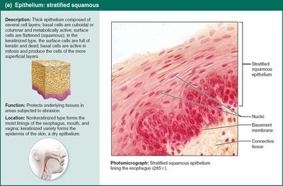
Stratified squamous epithelium: Protection; found in skin, esophagus.
Stratified cuboidal and columnar epithelium: Rare; found in sweat glands, pharynx.
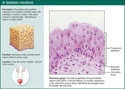
Transitional epithelium: Stretching; found in urinary bladder.
Glandular Epithelia
Gland: One or more cells that secrete an aqueous fluid (secretion).
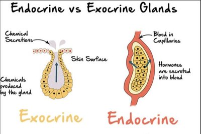
Endocrine glands: Ductless, release hormones into blood.
Exocrine glands: Release products onto body surfaces or into cavities.
Connective Tissue
Structure and Functions
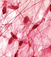
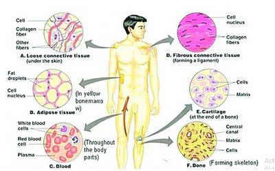
Connective tissue is the most abundant and widely distributed tissue in the body. It binds, supports, protects, insulates, stores fuel, and transports substances.
Four main classes: Connective tissue proper, Cartilage, Bone, Blood.
Major functions: Binding, support, protection, insulation, storage, transport.
Components of Connective Tissue
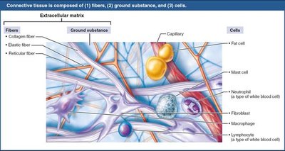
All connective tissues have three main components:
Ground substance: Fills space between cells and fibers.
Fibers: Collagen, elastic, and reticular fibers provide support.
Cells: Various cell types, including fibroblasts, chondroblasts, osteoblasts, and blood cells.
Connective Tissue Cells
Immature cells ("-blast"): Actively secrete matrix (e.g., fibroblasts, chondroblasts, osteoblasts).
Mature cells ("-cyte"): Maintain matrix (e.g., fibrocytes, chondrocytes, osteocytes).
Hematopoietic stem cells: Immature blood cells.
Other cell types: Adipocytes (fat storage), Leukocytes (immune defense), Mast cells (inflammation), Macrophages (phagocytosis).
Connective Tissue Categories
Connective tissue proper: Loose (areolar, adipose, reticular) and dense (regular, irregular, elastic).
Cartilage: Hyaline, elastic, fibrocartilage.
Bone: Compact and spongy bone.
Blood: Fluid tissue for transport.
Muscular Tissue
Structure and Functions
Muscular tissue consists of elongated cells specialized for contraction, enabling movement and force generation.
Skeletal muscle: Voluntary movement, attached to bones.
Smooth muscle: Involuntary, found in walls of hollow organs.
Cardiac muscle: Involuntary, found in heart.
Nervous Tissue
Structure and Functions
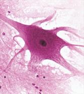
Nervous tissue is specialized for internal communication via electrical impulses. It consists of neurons and supporting glial cells.
Neurons: Transmit electrical signals.
Glial cells: Support and protect neurons.
Summary Table: Primary Tissue Classes
Tissue Type | Main Function | Key Locations |
|---|---|---|
Epithelial | Protection, secretion, absorption | Skin, lining of organs, glands |
Connective | Support, binding, transport | Bone, cartilage, blood, fat |
Muscular | Movement, force generation | Skeletal muscles, heart, digestive tract |
Nervous | Communication, control | Brain, spinal cord, nerves |
Additional info:
Histology is foundational for understanding organ structure and function in all chapters of Anatomy & Physiology.
Clinical relevance: Tissue pathology is central to disease diagnosis and treatment.
