Indietro
Chapter 9
Guida di studio - Note intelligenti
 Appunti personalizzati basati sui tuoi materiali, ampliati con definizioni chiave, esempi e contesto.
Joints (Articulations)
Introduction to Joints
Joints, or articulations, are the locations where two bones meet and where body movement occurs. The structure of a joint determines its function, with a tradeoff between strength and mobility. Stronger joints tend to be less mobile, while more mobile joints are generally weaker.
Classification of Joints
Structural and Functional Classifications
Structural classifications: Based on the material binding bones together and the presence of a joint cavity.
Fibrous – Bones joined by dense connective tissue; no joint cavity.
Cartilaginous – Bones joined by cartilage; no joint cavity.
Bony – Bones fuse together; no joint cavity.
Synovial – Bones separated by a fluid-filled joint cavity.
Functional classifications: Based on the amount of movement allowed.
Synarthrosis – Immovable joint (e.g., sutures of the skull).
Amphiarthrosis – Slightly movable joint (e.g., intervertebral discs).
Diarthrosis – Freely movable joint (e.g., most limb joints).
Types of Synarthroses (Immovable Joints)
Suture: Found only in the skull; bones interlock and are bound by dense fibrous connective tissue.
Gomphosis: Binds teeth to bony sockets via the periodontal ligament.
Synchondrosis: Cartilaginous bridge between two bones (e.g., epiphyseal plates, rib-sternum junctions).
Synostosis: Fusion of two bones (e.g., frontal bone, epiphyseal lines).
Types of Amphiarthroses (Slightly Movable Joints)
Syndesmosis: Bones connected by a ligament (e.g., distal tibiofibular joint).
Symphysis: Bones connected by fibrocartilage (e.g., pubic symphysis, intervertebral discs).
Synovial Joints (Diarthroses)
Basic Structure of Synovial Joints
Synovial joints are freely movable and are found at the ends of long bones. They are characterized by a joint cavity filled with synovial fluid, surrounded by an articular capsule, and covered by articular cartilage.
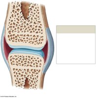
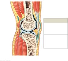
Articular cartilage: Covers articulating surfaces, preventing direct bone contact.
Joint (articular) capsule: Encloses the joint cavity and is lined by the synovial membrane.
Synovial fluid: Lubricates, distributes nutrients, and absorbs shock.
Accessory structures: Include menisci, fat pads, ligaments, tendons, and bursae.
Accessory Structures of Synovial Joints
Meniscus: Fibrocartilage pad that improves fit and absorbs shock.
Fat pads: Protect articular cartilages and fill spaces.
Ligaments: Support and strengthen joints; sprains occur when ligaments are stretched or torn.
Tendons: Attach muscles to bones, stabilizing joints.
Bursae: Fluid-filled sacs that reduce friction where tendons or ligaments rub against other tissues.
Stabilization of Synovial Joints
Stabilized by the shape of articulating surfaces, menisci, ligaments, tendons, and other bones or fat pads.
Stabilization prevents injury by limiting range of motion.
Movements at Synovial Joints
Planes and Types of Movement
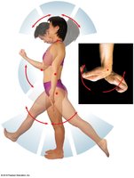
Movements are described by the plane or direction and the relationship between structures. Joints may be monaxial (one plane), biaxial (two planes), or triaxial (three planes).
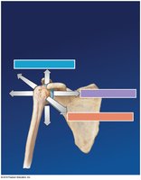
Gliding: Flat surfaces slide past each other (e.g., carpal bones).
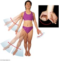
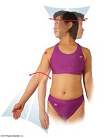
Angular movements: Change the angle between bones (flexion, extension, abduction, adduction, circumduction).
Rotation: Bone turns around its own long axis (medial/lateral rotation, pronation/supination).
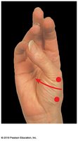
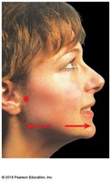
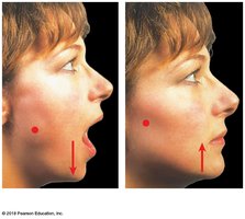
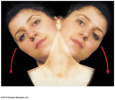
Special movements: Inversion, eversion, dorsiflexion, plantar flexion, opposition, reposition, protraction, retraction, elevation, depression, lateral flexion.
Examples of Joint Movements
Flexion/Extension/Hyperextension: Decreasing/increasing the angle between bones; hyperextension is extension beyond anatomical position.
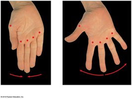
Abduction/Adduction: Movement away from/toward the midline.
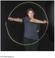
Circumduction: Circular movement combining flexion, extension, abduction, and adduction.
Rotation: Turning a bone around its long axis (e.g., head, limb rotation).
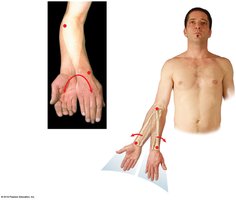
Pronation/Supination: Rotational movement of the forearm (palm down/up).
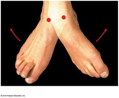
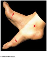
Inversion/Eversion: Twisting the sole of the foot medially/laterally.
Dorsiflexion/Plantar flexion: Flexion/extension at the ankle.
Opposition/Reposition: Movement of thumb toward/away from fingers.
Protraction/Retraction: Anterior/posterior movement in the horizontal plane.
Elevation/Depression: Moving a structure superiorly/inferiorly.
Lateral flexion: Bending the vertebral column to the side.
Types of Synovial Joints
Classification by Shape and Movement
Plane (gliding) joint: Flat or slightly curved surfaces; slight nonaxial movement (e.g., intercarpal joints).

Hinge joint: Angular motion in one plane (monaxial), like a door hinge (e.g., elbow, knee).

Condylar (ellipsoid) joint: Oval articular face within a depression; biaxial movement (e.g., wrist).

Saddle joint: Articular faces fit together like a rider in a saddle; biaxial (e.g., thumb carpometacarpal joint).

Pivot joint: Rotation only (monaxial) (e.g., proximal radioulnar joint, atlanto-axial joint).

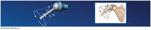
Ball-and-socket joint: Round head in a cup-shaped depression; triaxial movement (e.g., shoulder, hip).
Intervertebral Joints
Structure and Function
Intervertebral joints connect vertebrae and allow for flexibility and movement of the vertebral column. The first two cervical vertebrae are joined by a synovial joint, while adjacent vertebral bodies are connected by symphyses (intervertebral discs).
Intervertebral disc: Separates vertebral bodies; consists of anulus fibrosus (outer fibrocartilage) and nucleus pulposus (inner gelatinous core).
Vertebral end plates: Cartilage covering the superior and inferior surfaces of the disc.
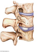
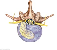
Bulging disc: Anulus fibrosus bulges into the vertebral canal.
Herniated disc: Nucleus pulposus breaks through anulus fibrosus, compressing spinal nerves.
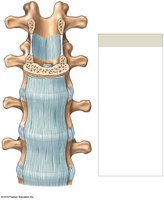
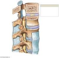
Intervertebral ligaments: Bind vertebrae together and stabilize the column (ligamenta flava, anterior/posterior longitudinal ligaments, interspinous, supraspinous ligaments).
Elbow and Knee Joints
Elbow Joint
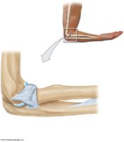
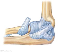
The elbow is a hinge joint involving the humerus, radius, and ulna. It allows flexion and extension with limited movement in other directions.
Humero-ulnar joint: Main articulation, between trochlea of humerus and trochlear notch of ulna.
Humeroradial joint: Between capitulum of humerus and head of radius.
Ligaments: Radial collateral, ulnar collateral, and anular ligaments stabilize the joint.
Knee Joint
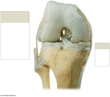
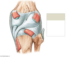
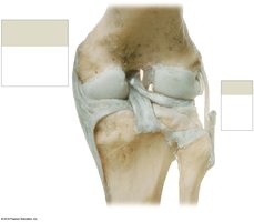
The knee is a complex hinge joint that transfers weight from the femur to the tibia. It consists of three articulations and is stabilized by menisci and multiple ligaments.
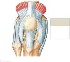
Articulations: Two femur-tibia (medial and lateral condyles), one femur-patella.
Menisci: Fibrocartilage pads that cushion and stabilize the joint.
Major ligaments: Patellar, popliteal, anterior/posterior cruciate, tibial collateral, fibular collateral.
Shoulder and Hip Joints
Shoulder Joint (Glenohumeral Joint)
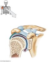
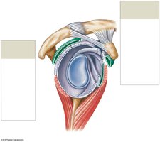
The shoulder is a ball-and-socket diarthrosis between the head of the humerus and the glenoid cavity of the scapula. It has the greatest range of motion but is the most frequently dislocated joint.
Glenoid labrum: Fibrocartilage rim that deepens the socket.
Ligaments: Acromioclavicular, coracoclavicular, coraco-acromial, coracohumeral, glenohumeral.
Muscles of the rotator cuff: Supraspinatus, infraspinatus, teres minor, subscapularis.
Bursae: Subdeltoid, subcoracoid, subacromial, subscapular.
Hip Joint
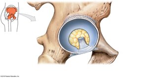
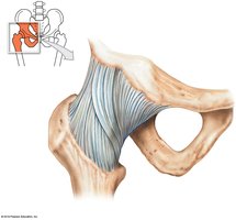
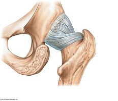
The hip is a strong ball-and-socket diarthrosis between the head of the femur and the acetabulum of the hip bone. It allows a wide range of motion and is stabilized by several ligaments and the acetabular labrum.
Acetabular labrum: Fibrocartilage rim that increases depth and seals in synovial fluid.
Ligaments: Iliofemoral, pubofemoral, ischiofemoral, transverse acetabular, ligament of the femoral head.
Effects of Aging on Joints
Degenerative Changes and Clinical Problems
Rheumatism: General pain and stiffness in the musculoskeletal system.
Arthritis: Inflammation of joints; includes osteoarthritis (wear and tear), rheumatoid arthritis (autoimmune), and gouty arthritis (uric acid crystals).
Joint immobilization reduces synovial fluid flow, increasing arthritis risk; treated by continuous passive motion (CPM).
Aging decreases bone mass, weakens bones, and increases fracture risk.
Integration with Other Body Systems
Functional Relationships
Bones undergo continuous remodeling (formation by osteoblasts, recycling by osteoclasts).
Balance is affected by age, physical stress, hormones, mineral uptake/excretion, and genetics.
Muscles attach to bones for movement; endocrine, digestive, and urinary systems regulate mineral supply.
The skeleton serves as a mineral reserve and supports other organ systems.