 Indietro
Neurons: Cellular and Network Properties – Study Notes
Guida di studio - Note intelligenti
 Appunti personalizzati basati sui tuoi materiali, ampliati con definizioni chiave, esempi e contesto.
Neurons: Cellular and Network Properties
Overview of the Nervous System
The nervous system is a complex network responsible for coordinating body functions and enabling communication between different body regions. It is divided into the central nervous system (CNS) and peripheral nervous system (PNS), each with specialized roles and cellular components.
Central Nervous System (CNS): Consists of the brain and spinal cord; integrates and processes information.
Peripheral Nervous System (PNS): Includes all neural tissue outside the CNS; subdivided into sensory (afferent) and motor (efferent) divisions.
Enteric Nervous System: A network of neurons in the digestive tract, capable of autonomous function but regulated by the autonomic nervous system.
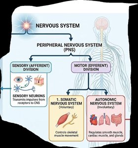
Divisions of the Peripheral Nervous System
The PNS is further divided based on function:
Sensory (Afferent) Division: Transmits impulses from sensory receptors to the CNS.
Motor (Efferent) Division: Transmits commands from the CNS to effector organs. It includes:
Somatic Nervous System: Controls voluntary skeletal muscle movement.
Autonomic Nervous System (ANS): Regulates involuntary functions (smooth muscle, cardiac muscle, glands).
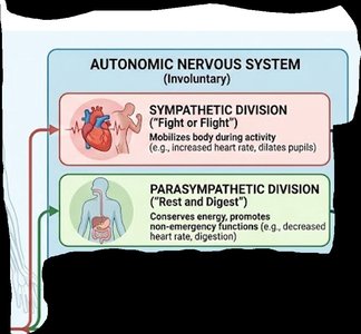
Autonomic Nervous System Subdivisions
Sympathetic Division: Mobilizes the body for activity ("fight or flight"), increasing heart rate and dilating pupils.
Parasympathetic Division: Conserves energy and promotes non-emergency functions ("rest and digest"), such as decreasing heart rate and stimulating digestion.
Cells of the Nervous System
Neurons: Structure and Function
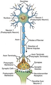
Neurons are the functional units of the nervous system, specialized for the transmission of electrical and chemical signals. They are classified by structure and function:
Cell Body (Soma): Contains the nucleus and organelles; metabolic center of the neuron.
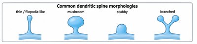
Dendrites: Receive incoming signals from other neurons; may have specialized protrusions called dendritic spines that increase surface area for synaptic input.
Axon: Conducts electrical impulses away from the cell body toward other neurons or effectors; may be myelinated for faster signal transmission.
Axon Terminals: Release neurotransmitters into the synaptic cleft to communicate with postsynaptic cells.
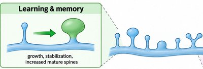
Dendritic Spines and Their Role
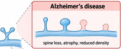
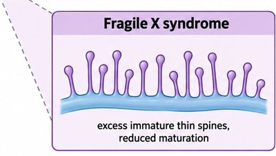
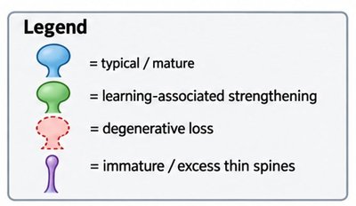
Dendritic spines are small protrusions on dendrites that serve as the primary sites of synaptic input. Their shape and density are associated with learning, memory, and certain neurological disorders.
Learning & Memory: Growth and stabilization of mature spines are linked to synaptic strengthening.
Alzheimer's Disease: Characterized by spine loss, atrophy, and reduced density.
Fragile X Syndrome: Marked by excess immature thin spines and reduced maturation.
Functional Categories of Neurons
Neurons are classified by their function and structure:
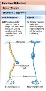
Sensory (Afferent) Neurons: Transmit sensory information to the CNS.
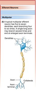
Efferent Neurons: Carry commands from the CNS to muscles and glands.
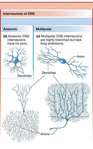
Interneurons: Connect neurons within the CNS; may be anaxonic (no axon) or multipolar (highly branched).
Axonal Transport
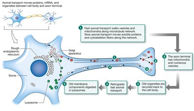
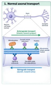
Axonal transport is the process by which materials are moved between the cell body and axon terminals. It is essential for neuron function and survival.
Fast Axonal Transport: Moves organelles and vesicles rapidly (up to 400 mm/day) using motor proteins along microtubules.
Slow Axonal Transport: Moves cytoskeletal and soluble proteins more slowly (0.2–2.5 mm/day).
Anterograde Transport: From cell body to axon terminal (kinesin motor protein).
Retrograde Transport: From axon terminal to cell body (dynein motor protein).
Pathologies: Alzheimer's Disease and Axonal Transport
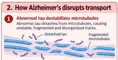
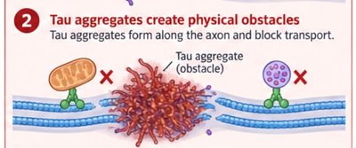
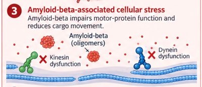
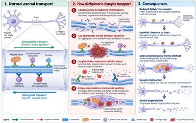
Alzheimer's disease disrupts axonal transport through several mechanisms:
Abnormal Tau Protein: Hyperphosphorylated tau detaches from microtubules, destabilizing tracks and causing fragmentation.
Tau Aggregates: Form physical obstacles along axons, blocking transport.
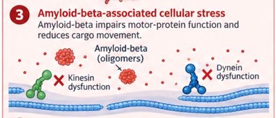
Amyloid-Beta Oligomers: Impair motor protein function, reducing cargo movement.
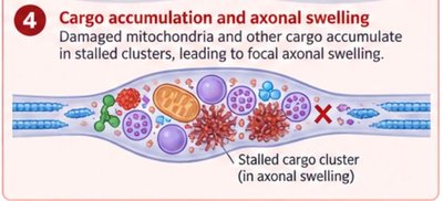
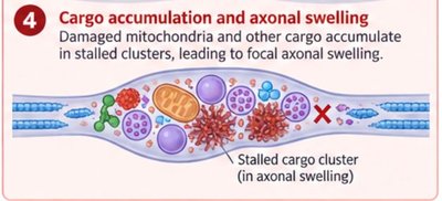
Cargo Accumulation: Damaged mitochondria and other cargo accumulate, leading to axonal swelling and degeneration.
Glial Cells and Their Functions
Glial cells provide structural and functional support for neurons. They are found in both the CNS and PNS and have diverse roles.
Schwann Cells (PNS) and Oligodendrocytes (CNS): Form myelin sheaths around axons, increasing conduction speed; nodes of Ranvier are gaps in myelin.
Astrocytes (CNS): Regulate the extracellular environment, form the blood-brain barrier, and provide metabolic support.
Microglia (CNS): Act as immune cells, removing debris and pathogens.
Ependymal Cells (CNS): Line ventricles, create barriers, and serve as a source of neural stem cells.

Neural Damage and Regeneration
Neural injury responses differ between the CNS and PNS:
Cell Body Damage: Results in neuron death.
Axon Severing: The cell body and attached segment may survive; the distal segment degenerates.
PNS Regeneration: Schwann cells and macrophages clear debris, and surviving axons regrow through Schwann cell tubes toward targets.
CNS Regeneration: Less likely due to glial scarring and inhibitory environment.
Electrical Signals in Neurons
Membrane Potential and Ion Movement
Neurons are excitable cells capable of generating and propagating electrical signals. These signals are based on changes in membrane potential, which is determined by ion gradients and membrane permeability.
Nernst Equation: Predicts the equilibrium potential for a single ion:
Goldman-Hodgkin-Katz (GHK) Equation: Calculates membrane potential considering multiple ions and their permeabilities:
Key Ions: Potassium (K+), Sodium (Na+), Chloride (Cl-).
Resting Membrane Potential: Dominated by K+ due to high permeability via leak channels.
Ion Channels and Membrane Permeability
Ion channels are selective and control the permeability of the neuronal membrane. Types include:
Mechanically Gated Channels
Chemically Gated Channels
Voltage-Gated Channels
The size and charge of the channel pore determine ion selectivity.
Types of Electrical Signals
Graded Potentials: Variable strength, short-distance signals, occur in dendrites and cell body, can be summed, lose strength with distance.
Action Potentials: All-or-none, long-distance signals, occur in axons, initiated at the trigger zone, cannot be summed, have a refractory period.
Graded Potential | Action Potential | |
|---|---|---|
Type of Signal | Input signal | Regenerating conduction signal |
Location | Dendrites, cell body | Trigger zone, axon |
Channels Involved | Mechanically, chemically, or voltage-gated | Voltage-gated |
Ions Involved | Na+, K+, Ca2+, Cl- | Na+, K+ |
Signal Strength | Variable, can be summed | All-or-none, cannot be summed |
Initiation | Entry of ions through channels | Above-threshold graded potential at trigger zone |
Unique Features | No minimum level, summation possible | Threshold required, refractory period |
Graded Potentials and Signal Propagation
Graded potentials are local changes in membrane potential that decrease in strength as they spread due to current leak and cytoplasmic resistance. If strong enough, they reach the axon hillock and trigger an action potential.
Excitatory Graded Potentials: Depolarize the membrane, increasing the likelihood of action potential firing.
Inhibitory Graded Potentials: Hyperpolarize the membrane, decreasing the likelihood of action potential firing.
Cell Excitability: The ability of a neuron to fire an action potential in response to a stimulus.
