 Indietro
Respiratory Physiology: Lecture 2
Guida di studio - Note intelligenti
 Appunti personalizzati basati sui tuoi materiali, ampliati con definizioni chiave, esempi e contesto.
The Respiratory System
Overview of the Respiratory Tract
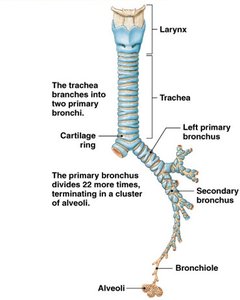
The respiratory system is responsible for the exchange of gases (oxygen and carbon dioxide) between the body and the environment. It consists of a series of branching tubes that conduct air to the alveoli, where gas exchange occurs.
Larynx: The uppermost part of the lower respiratory tract, involved in sound production and airway protection.
Trachea: A rigid tube supported by cartilage rings, conducting air from the larynx to the bronchi.
Primary Bronchi: The trachea divides into left and right primary bronchi, each entering a lung.
Bronchial Tree: The bronchi branch repeatedly (up to 22 times) into smaller bronchioles, ending in clusters of alveoli.
Microscopic Structure of the Lungs
Lung Lobule and Alveolar Organization
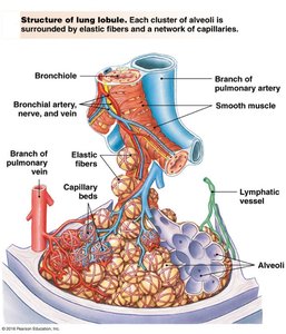
Each lung is divided into lobules, which are the smallest functional units of the lung. Each lobule contains clusters of alveoli, surrounded by a network of capillaries and elastic fibers.
Bronchioles: Small airways lacking cartilage, leading to alveolar ducts and alveoli.
Alveoli: Tiny air sacs where gas exchange occurs. Each alveolus is surrounded by capillary beds and elastic fibers.
Capillary Beds: Dense networks of capillaries facilitate efficient gas exchange.
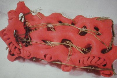
Elastic Fibers: Provide recoil necessary for expiration and help maintain alveolar structure.
Lymphatic Vessels: Drain excess fluid and participate in immune defense.
Alveolar Structure and Gas Exchange
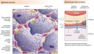
The alveolar wall is extremely thin, optimizing it for rapid gas exchange. It consists of several cell types and is closely associated with capillaries.
Type I Alveolar Cells: Flat cells forming the majority of the alveolar surface, specialized for gas exchange.
Type II Alveolar Cells: Secrete surfactant, reducing surface tension and preventing alveolar collapse.
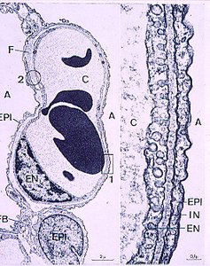
Capillary Endothelium: Forms the other side of the respiratory membrane, allowing gases to diffuse between air and blood.
Respiratory Membrane: Composed of alveolar epithelium, fused basement membrane, and capillary endothelium; typically 0.1–1.5 μm thick.
Histological and Ultrastructural Views of the Alveoli
Microscopic and Electron Micrographs
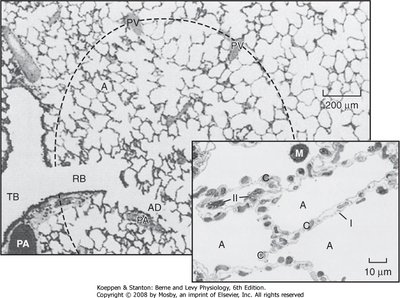
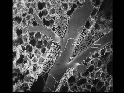
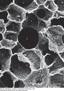
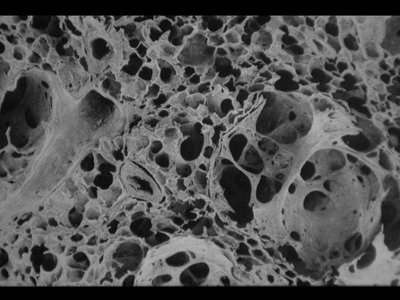
Histological and electron micrographs reveal the intricate structure of alveoli and their relationship with pulmonary vessels.
Alveolar Sacs: Clusters of alveoli sharing common walls, maximizing surface area for gas exchange.
Pulmonary Arteries and Veins: Closely associated with alveoli, facilitating efficient oxygen uptake and carbon dioxide removal.
Interalveolar Septa: Thin walls between adjacent alveoli, containing capillaries and connective tissue.
Alveolar Mechanics and Elastic Properties
Elastic Fibers and Alveolar Interdependence
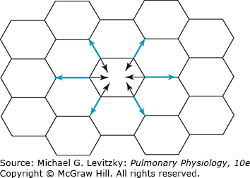
Elastic fibers in the lung parenchyma provide the recoil necessary for expiration and help maintain the structural integrity of the alveoli. The interconnected nature of alveoli means that changes in one alveolus affect its neighbors, a concept known as alveolar interdependence.
Elastic Recoil: The tendency of the lungs to return to their original size after being stretched.
Alveolar Interdependence: The structural support provided by adjacent alveoli prevents collapse and maintains uniform inflation.
Mechanics of Breathing
Boyle's Law and Pulmonary Ventilation
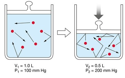
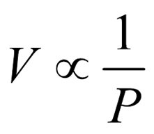
Pulmonary ventilation is governed by Boyle's Law, which states that the pressure of a gas is inversely proportional to its volume at constant temperature. This principle explains how changes in thoracic volume drive airflow into and out of the lungs.
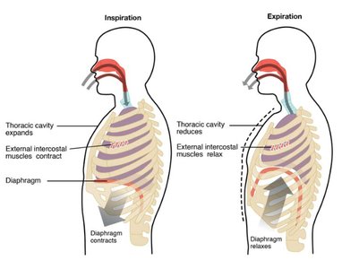
Inspiration: Thoracic cavity volume increases, intrapulmonary pressure decreases, and air flows into the lungs.
Expiration: Thoracic cavity volume decreases, intrapulmonary pressure increases, and air flows out of the lungs.
Muscles of Respiration
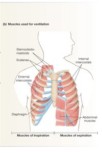
Breathing involves the coordinated action of several muscle groups:
Diaphragm: The primary muscle of inspiration; contraction increases thoracic volume.
External Intercostals: Elevate the ribs during inspiration, expanding the thoracic cavity.
Internal Intercostals and Abdominal Muscles: Active during forced expiration, decreasing thoracic volume.
Summary Table: Key Structures and Functions in Pulmonary Anatomy
Structure | Function |
|---|---|
Trachea | Conducts air to bronchi; supported by cartilage rings |
Bronchi/Bronchioles | Branching airways leading to alveoli |
Alveoli | Site of gas exchange |
Capillaries | Transport blood for gas exchange |
Elastic Fibers | Provide recoil and maintain structure |
Diaphragm | Main muscle of inspiration |
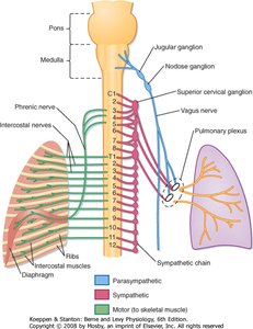
Intercostal Muscles | Assist in expanding and compressing the thoracic cavity |
Key Equations
Boyle's Law:
Where is volume and is pressure. As volume increases, pressure decreases, and vice versa.
