 Indietro
The Cardiovascular System: The Heart – Structure, Function, and Physiology
Guida di studio - Note intelligenti
 Appunti personalizzati basati sui tuoi materiali, ampliati con definizioni chiave, esempi e contesto.
The Cardiovascular System: The Heart
Location and Basic Structure of the Heart
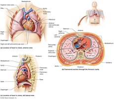
The heart is a muscular organ responsible for pumping blood throughout the body. It is located in the mediastinum, a subdivision of the thoracic cavity, and sits slightly to the left within the pericardial cavity. The heart is cone-shaped, with the apex pointing toward the left hip and the base facing the posterior rib cage. It is about the size of a fist and weighs between 250–350 grams.
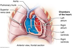
Chambers: Four hollow chambers: right and left atria (superior), right and left ventricles (inferior).
Sulci: The atrioventricular sulcus separates atria from ventricles; the interventricular sulcus separates the right and left ventricles.
Major Blood Vessels and Blood Flow
Veins: Superior and inferior venae cavae deliver deoxygenated blood to the right atrium; pulmonary veins deliver oxygenated blood to the left atrium.
Arteries: Right ventricle pumps blood into the pulmonary trunk; left ventricle pumps blood into the aorta.
Great Vessels: Collectively refers to the main veins and arteries entering and leaving the heart.
Circuits of Blood Flow
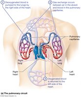
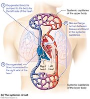
The heart functions as two pumps, each serving a different circuit:
Pulmonary Circuit: Right side pumps deoxygenated blood to the lungs for gas exchange (low-pressure circuit).
Systemic Circuit: Left side pumps oxygenated blood to the body (high-pressure circuit).
Functions of the Heart
Maintains homeostasis of blood pressure by regulating the rate and force of contractions.
Acts as an endocrine organ by producing atrial natriuretic peptide (ANP), which lowers blood pressure by decreasing sodium and water retention in the kidneys.
The Pericardium, Heart Wall, and Heart Skeleton
Pericardium
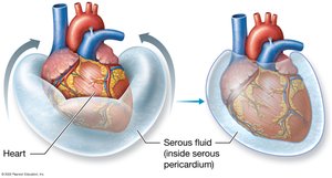
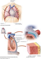
The pericardium is a double-layered sac surrounding the heart:
Fibrous Pericardium: Tough outer layer that anchors the heart and prevents overexpansion.
Serous Pericardium: Thin inner layer with two parts: parietal (lines the cavity) and visceral (epicardium, adheres to the heart).
Pericardial Cavity: Space between layers, filled with serous fluid to reduce friction.
Heart Wall and Skeleton
Epicardium (Visceral Pericardium): Outermost layer.
Myocardium: Thick middle layer of cardiac muscle cells (myocytes) arranged in a spiral pattern; responsible for contraction.
Fibrous Skeleton: Dense connective tissue providing structural support and electrical insulation.
Endocardium: Innermost layer of endothelial cells and connective tissue, forming a blood-heart barrier.
Clinical Connections
Thoracotomy: Surgical opening of the thoracic cavity for access to the heart and lungs.
Cardiac Tamponade: Accumulation of excess fluid in the pericardial cavity, compressing the heart and reducing cardiac output.
Myocarditis: Inflammation of the myocardium, often due to viral infection (e.g., SARS-CoV-2).
The Great Vessels, Chambers, and Valves of the Heart
The Great Vessels
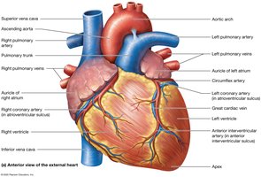
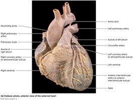
Superior/Inferior Vena Cava: Drain deoxygenated blood into the right atrium.
Pulmonary Trunk: Receives blood from the right ventricle, splits into right and left pulmonary arteries.
Pulmonary Veins: Four veins return oxygenated blood from the lungs to the left atrium.
Aorta: Largest artery, carries oxygenated blood from the left ventricle to the body.
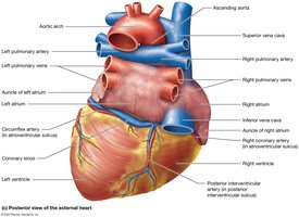
Chambers of the Heart
Atria: Right atrium (large, thin-walled, anterior); left atrium (smaller, thicker-walled, posterior). Both have auricles for expansion.
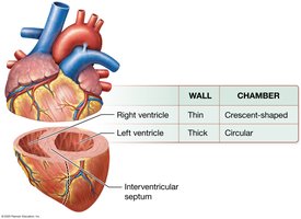
Ventricles: Right ventricle (wider, thinner walls); left ventricle (thicker walls, pumps against higher resistance).
Internal Features: Pectinate muscles (right atrium), trabeculae carneae (ventricles), papillary muscles, chordae tendineae, interventricular septum.
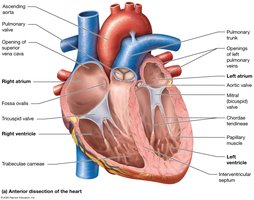
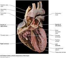
Valves of the Heart
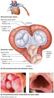
Atrioventricular (AV) Valves: Tricuspid (right), bicuspid/mitral (left); prevent backflow into atria, supported by chordae tendineae and papillary muscles.
Semilunar (SL) Valves: Pulmonary (right ventricle to pulmonary trunk), aortic (left ventricle to aorta); prevent backflow into ventricles.
Valvular Heart Diseases
Insufficient Valve: Fails to close fully, causing regurgitation.
Stenotic Valve: Stiffened by calcium deposits, impeding blood flow.
Heart Murmur: Audible sound due to turbulent blood flow.

Blood Flow Through the Heart
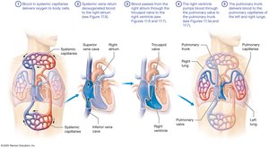
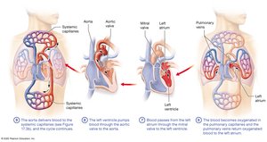
Blood flows in a specific sequence through the heart chambers and valves, ensuring unidirectional movement and efficient oxygenation.
The Coronary Circulation
Coronary Arteries
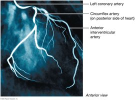
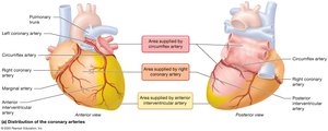
Right Coronary Artery: Branches into marginal and posterior interventricular arteries.
Left Coronary Artery: Branches into anterior interventricular (LAD) and circumflex arteries.
Anastomoses: Alternate routes for blood flow, providing collateral circulation.
Coronary Veins
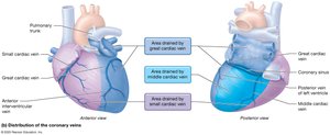
Coronary Sinus: Main vein draining into the right atrium.
Great, Small, and Middle Cardiac Veins: Drain different regions of the heart.
Coronary Artery Disease and Myocardial Infarction
Coronary Artery Disease (CAD): Buildup of plaques reduces blood flow, causing ischemia and angina pectoris.
Myocardial Infarction (MI): Heart attack due to blocked artery, resulting in death of cardiac muscle cells.
Treatments: Lifestyle changes, medications, angioplasty, stent placement, or coronary artery bypass grafting.
Cardiac Muscle Tissue Anatomy and Electrophysiology
Histology of Cardiac Muscle Tissue
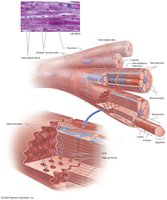
Striations: Due to arrangement of contractile proteins.
Intercalated Discs: Contain desmosomes and gap junctions for mechanical and electrical connection, allowing the heart to function as a functional syncytium.
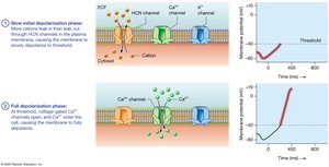
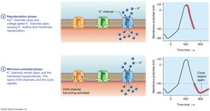
Ion Channels: Voltage-gated sodium, calcium, potassium, and HCN channels regulate action potentials.
Electrophysiology: Pacemaker Cells and the Cardiac Conduction System
Pacemaker Cells: Generate spontaneous action potentials, setting the heart's rhythm (autorhythmicity).
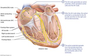
Conduction System: Includes SA node (primary pacemaker), AV node, and Purkinje fiber system (AV bundle, bundle branches, terminal branches).
Sinus Rhythm: Normal rhythm set by the SA node.
Ectopic Pacemaker: Other cells attempt to pace the heart, causing arrhythmias.
Electrophysiology: Contractile Cells
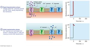
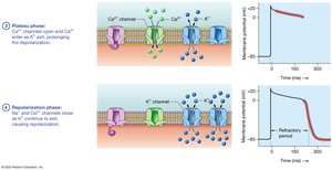
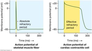
Action Potential Phases: Rapid depolarization (Na+ influx), initial repolarization (K+ outflow), plateau (Ca2+ influx balances K+ outflow), repolarization (K+ outflow).
Plateau Phase: Prolongs action potential, prevents tetany, and allows for effective refractory period.
Excitation-Contraction Coupling: Involves Ca2+-induced Ca2+ release from the sarcoplasmic reticulum.
The Electrocardiogram (ECG/EKG)
P Wave: Atrial depolarization.
QRS Complex: Ventricular depolarization (and atrial repolarization).
T Wave: Ventricular repolarization.
Intervals and Segments: R-R (heart rate), P-R (atrial to ventricular conduction), Q-T (ventricular action potentials), S-T (plateau phase).
Dysrhythmias
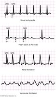
Bradycardia: Heart rate < 60 bpm.
Tachycardia: Heart rate > 100 bpm.
Heart Block: Conduction pathway disturbances, often at the AV node.
Fibrillation: Disorganized electrical activity; atrial fibrillation is less dangerous, ventricular fibrillation is life-threatening.




Mechanical Physiology of the Heart: The Cardiac Cycle
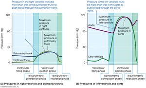
Cardiac Cycle Phases
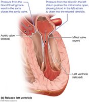
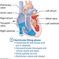
Diastole: Relaxation phase; chambers fill with blood.
Systole: Contraction phase; blood is ejected.
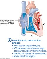
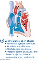
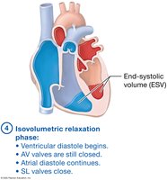
Four Main Phases:
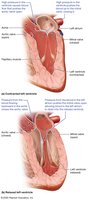
Ventricular filling (AV valves open, SL valves closed)
Isovolumetric contraction (all valves closed)
Ventricular ejection (SL valves open, AV valves closed)
Isovolumetric relaxation (all valves closed)
Heart Sounds
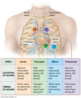
S1 (Lub): Closure of AV valves.
S2 (Dub): Closure of SL valves.
Heart Murmurs: Abnormal sounds due to turbulent flow, often from defective valves.
Extra Heart Sounds: S3 and S4, may indicate pathology.

Cardiac Output and Regulation
Heart Rate (HR): Beats per minute (normal: 60–80 bpm).
Stroke Volume (SV): Blood pumped per beat.
Cardiac Output (CO): Blood pumped per minute;
Stroke Volume Calculation: (End-Diastolic Volume minus End-Systolic Volume)
Factors Influencing Stroke Volume
Preload: Degree of stretch before contraction (Frank-Starling Law).
Contractility: Force of contraction at a given preload (influenced by inotropic agents).
Afterload: Pressure the heart must overcome to eject blood.
Ventricular Hypertrophy
Enlargement of ventricles due to chronic increased afterload (e.g., hypertension).
Decreases chamber volume, reducing filling and output.
Regulation of Cardiac Output
Autonomic Nervous System: Sympathetic stimulation increases HR and contractility; parasympathetic (vagus nerve) decreases HR.
Endocrine System: Epinephrine, norepinephrine, thyroid hormone, glucagon, aldosterone, and ADH affect HR and SV.6
Other Factors: Electrolyte concentrations, body temperature, age, and physical fitness.
Heart Failure
Condition where the heart cannot pump effectively.
Left Ventricular Failure: Leads to pulmonary congestion and edema.
Right Ventricular Failure: Causes systemic congestion and peripheral edema.
Treatment: Lifestyle changes, medications, and sometimes surgery to improve cardiac output and reduce fluid overload.
Summary Table: Key Structures and Functions of the Heart
Structure | Function |
|---|---|
Atria | Receive blood returning to the heart |
Ventricles | Pump blood out of the heart |
Valves (AV & SL) | Ensure unidirectional blood flow |
Coronary Vessels | Supply blood to the heart muscle |
Conduction System | Coordinates heartbeat |
Additional info: This guide integrates foundational concepts in cardiac anatomy and physiology, including clinical correlations and regulatory mechanisms, to provide a comprehensive overview suitable for exam preparation.
