 Indietro
The Integumentary System: Structure and Function of Skin and Its Appendages
Guida di studio - Note intelligenti
 Appunti personalizzati basati sui tuoi materiali, ampliati con definizioni chiave, esempi e contesto.
Integumentary System
Overview and Functions
The integumentary system consists of the skin and its accessory organs, including hair, nails, and cutaneous glands. As the largest organ system, the skin serves as a protective barrier and plays critical roles in homeostasis.
Barrier: Protects against mechanical injury, pathogens, and water loss.
Vitamin D Synthesis: Initiates the synthesis of vitamin D upon exposure to UV light.
Sensation: Contains sensory receptors for touch, pain, and temperature.
Thermoregulation: Regulates body temperature via sweat and blood flow.
Nonverbal Communication: Facial expressions and appearance convey social signals.
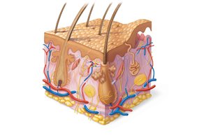
Structure of the Skin
Major Layers
The skin is composed of three main layers, each with distinct structures and functions:
Epidermis: Outermost, avascular layer of keratinized stratified squamous epithelium.
Dermis: Middle, vascularized layer of connective tissue containing hair follicles, glands, and sensory structures.
Hypodermis (Subcutaneous Tissue): Deepest layer, primarily areolar and adipose tissue, not technically part of the skin but anchors it to underlying structures.
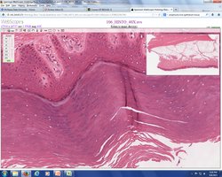
Epidermis
General Characteristics
The epidermis is a keratinized, stratified squamous epithelium composed mainly of keratinocytes. It is avascular and relies on diffusion from the underlying dermis for nutrients. The epidermis is constantly renewed by apoptosis and shedding of superficial cells.
Keratinocytes: Produce keratin, a protein that provides strength and waterproofing.
Melanocytes: Produce melanin, which protects against UV radiation.
Dendritic (Langerhans) Cells: Immune cells that detect pathogens.
Tactile (Merkel) Cells: Sensory receptors for touch.
Stem Cells: Located in the basal layer, responsible for regeneration.
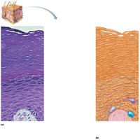
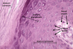
Layers of the Epidermis
The epidermis is organized into distinct layers (strata). Thick skin (palms, soles) has five layers; thin skin has four (lacking stratum lucidum).
Stratum Basale: Deepest layer; single row of mitotically active stem cells, melanocytes, and some dendritic cells.
Stratum Spinosum: Several layers of keratinocytes connected by desmosomes; contains dendritic cells and melanosomes.
Stratum Granulosum: 1–5 layers; keratinization begins, cells accumulate keratohyaline and lamellar granules (waterproofing glycolipids).
Stratum Lucidum: Only in thick skin; thin, translucent layer of dead keratinocytes.
Stratum Corneum: 20–30 layers of dead, anucleated keratinized cells; provides a durable overcoat.
Dermis
General Characteristics and Layers
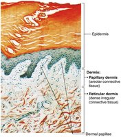
The dermis is a connective tissue layer that supports the epidermis and houses blood vessels, nerves, hair follicles, and glands. It is divided into two layers:
Papillary Layer: Superficial, areolar connective tissue with dermal papillae that interlock with the epidermis, increasing surface area for exchange and anchoring.
Reticular Layer: Deeper, dense irregular connective tissue providing strength and elasticity.
Dermal Modifications and Skin Markings
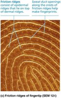
Friction Ridges: Form fingerprints; enhance grip and tactile sensitivity.
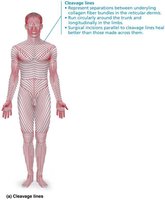
Cleavage (Tension) Lines: Orientation of collagen fibers; important for surgical incisions to minimize scarring.
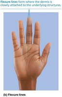
Flexure Lines: Dermal folds at joints; allow skin to move with underlying structures.
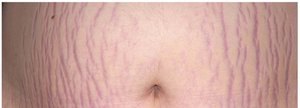
Stretch Marks (Striae): Tears in the dermis due to rapid stretching (e.g., pregnancy, obesity).
Hypodermis (Subcutaneous Tissue)
Structure and Function
The hypodermis is composed mainly of areolar and adipose tissue. It anchors the skin to underlying structures, provides insulation, and serves as an energy reserve.
Skin Color
Pigments and Clinical Significance
Skin color is determined by the amount and type of pigments present:
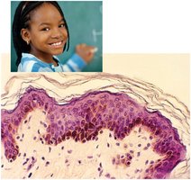
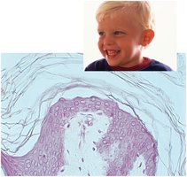
Melanin: Produced by melanocytes; eumelanin (brown-black) and pheomelanin (yellow-red).
Carotene: Yellow to orange pigment from diet, accumulates in stratum corneum and fat.
Hemoglobin: Red pigment in blood, visible in fair skin.
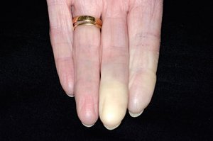
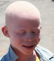
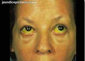
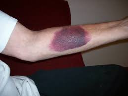
Disorders of skin color include cyanosis (bluish), erythema (redness), pallor (paleness), albinism (lack of melanin), jaundice (yellowing), and hematoma (bruising).




Hair
Structure and Function
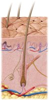
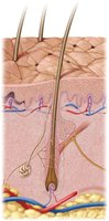
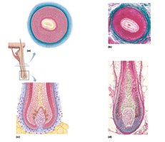
Hair is composed of dead, keratinized cells and serves various functions, including protection, sensation, and thermoregulation. The hair follicle is a complex structure extending from the epidermis into the dermis.
Shaft: Visible part above the skin surface.
Root: Portion within the skin.
Bulb: Expanded base where hair growth occurs.
Hair Matrix: Actively dividing cells producing the hair.
Dermal Papilla: Provides nutrients and signals for hair growth.
Arrector Pili Muscle: Smooth muscle causing "goosebumps."
Types of Hair
Lanugo: Fine, unpigmented hair on fetuses.
Vellus: Fine, pale hair on children and adult females.
Terminal Hair: Coarse, pigmented hair (scalp, eyebrows, eyelashes, axillary, and pubic regions).

Hair Growth and Loss
Hirsutism: Excessive hair growth, often due to hormonal imbalance.
Alopecia: Hair loss or baldness.
Nails
Structure and Function
Nails are clear, hard derivatives of the stratum corneum, composed of hard keratin. They protect the distal phalanx and enhance fine touch.
Nail Matrix: Growth zone under the proximal nail fold.
Lunule: White crescent at the base of the nail.
Hyponychium: Skin under the free edge of the nail.
Cutaneous Glands
Types and Functions
Sudoriferous (Sweat) Glands: Eccrine (merocrine) and apocrine types; involved in thermoregulation and scent communication.
Eccrine Glands: Widely distributed, secrete watery sweat for cooling.
Apocrine Glands: Found in axillary and genital areas; secrete milky sweat containing fatty acids and proteins.
Ceruminous Glands: Modified apocrine glands in the ear canal; produce earwax (cerumen).
Mammary Glands: Modified apocrine glands; produce milk.
Sebaceous Glands: Secrete oily sebum into hair follicles; lubricates and waterproofs skin and hair.
Skin Disorders
Skin Cancer
Skin cancer is primarily induced by ultraviolet (UV) radiation. The three main types are classified by the epidermal cell of origin:
Basal Cell Carcinoma: Most common, arises from stratum basale; rarely metastasizes.
Squamous Cell Carcinoma: Arises from keratinocytes of the stratum spinosum; can metastasize if untreated.
Malignant Melanoma: Arises from melanocytes; highly metastatic and most deadly.
Burns
Burns are classified by depth and extent of tissue damage:
First-Degree: Involves only the epidermis; redness and pain.
Second-Degree: Involves epidermis and upper dermis; blisters and pain.
Third-Degree: Destroys entire thickness of skin; requires grafting.
Additional info: The notes above integrate textbook-level explanations and expand on the provided slides for clarity and completeness, suitable for college-level Anatomy & Physiology students.
