 Indietro
The Integumentary System: Structure and Function of Skin and Its Appendages
Guida di studio - Note intelligenti
 Appunti personalizzati basati sui tuoi materiali, ampliati con definizioni chiave, esempi e contesto.
Integumentary System
Overview and Functions
The integumentary system consists of the skin and its accessory organs, including hair, nails, and cutaneous glands. The skin is the largest organ of the body and serves multiple essential functions:
Barrier: Protects against mechanical injury, pathogens, and water loss.
Vitamin D Synthesis: Initiates synthesis of vitamin D when exposed to sunlight.
Sensation: Contains sensory receptors for touch, pain, temperature, and pressure.
Thermoregulation: Regulates body temperature via sweat glands and blood vessels.
Nonverbal Communication: Expresses emotions through facial expressions and skin color changes.
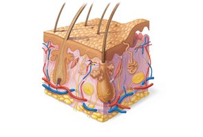
The skin is composed of three main layers: epidermis, dermis, and hypodermis (subcutaneous tissue).
Epidermis
General Characteristics
The epidermis is the outermost layer of skin, composed of keratinized stratified squamous epithelium. It is avascular (lacks blood vessels) and contains free nerve endings. The epidermis undergoes constant apoptosis, shedding approximately 50,000 cells every minute.
Cells of the Epidermis
Keratinocytes: Produce keratin, a protein that provides strength and waterproofing.
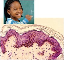
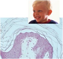
Melanocytes: Produce melanin, the pigment responsible for skin color.
Dendritic Cells: Macrophages involved in immune defense.
Tactile (Merkel) Cells: Sensory receptors for touch.
Stem Cells: Located in the basal layer, responsible for regeneration.
Layers of the Epidermis
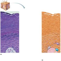
The epidermis is organized into four or five distinct layers, depending on skin thickness:
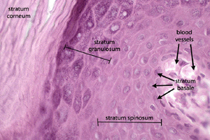
Stratum Basale: Deepest layer; contains actively mitotic stem cells and melanocytes.
Stratum Spinosum: Several layers of keratinocytes unified by desmosomes; contains dendritic cells.
Stratum Granulosum: Keratinization and waterproofing occur; cells contain keratohyaline and lamellar granules.
Stratum Lucidum: Present only in thick skin; consists of dead cells.
Stratum Corneum: Most superficial; 20–30 layers of anucleated, keratinized dead cells.
Waterproofing the Skin
Waterproofing is primarily achieved in the stratum granulosum through the production of keratohyaline granules (containing filaggrin protein, which binds keratin) and lamellar granules (which release glycolipids).
Dermis
General Characteristics
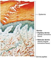
The dermis is a connective tissue layer beneath the epidermis. It is vascularized and contains hair follicles, cutaneous glands, and sensory nerve structures. The dermis is divided into two layers:
Papillary Layer: Areolar connective tissue; contains dermal papillae, capillary loops, and touch receptors.
Reticular Layer: Dense irregular connective tissue; provides strength and elasticity.
Dermal Modifications and Skin Markings
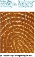
Friction Ridges: Formed by dermal papillae and epidermal ridges; responsible for fingerprints.
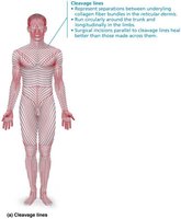
Cleavage Lines: Collagen fibers run parallel to skin surface; important for surgical incisions.
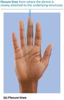
Flexure Lines: Dermal folds at or near joints; tightly secured to deeper structures, causing deep creases.
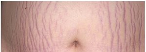
Stretch Marks (Striae): Result from dermal tears due to rapid stretching.
Hypodermis (Subcutaneous Tissue)
Structure and Function
The hypodermis lies beneath the dermis and is not considered part of the skin. It consists of areolar and adipose tissue, providing insulation, energy storage, and cushioning.
Skin Color
Pigments and Disorders
Skin color is determined by several pigments:
Melanin: Produced by melanocytes; includes eumelanin (brown-black) and pheomelanin (yellow-red).
Carotene: Yellow to orange pigment from diet.
Hemoglobin: Red pigment in blood.
Disorders of skin color include:
Cyanosis: Bluish discoloration due to lack of oxygen.
Erythema: Redness from increased blood flow.
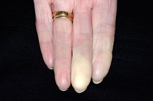
Pallor: Paleness from reduced blood flow.
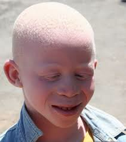
Albinism: Lack of melanin production.
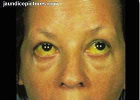
Jaundice: Yellowing from bilirubin accumulation.
Hematoma: Bruising from blood accumulation under skin.




Hair
Structure and Function
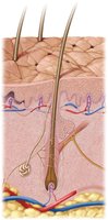
Hair is composed of dead, keratinized cells and is divided into three zones: shaft, root, and bulb. The hair follicle contains the hair matrix (site of growth), dermal papilla (provides nutrients), and melanocytes (pigmentation). Functions of hair include warning of insects, protection from physical trauma, reduction of heat loss, and shielding from sunlight.
Hair Types
Lanugo: Fine, downy, unpigmented hair (fetal).
Vellus: Fine, pale hair (children and adult females).
Terminal Hair: Longer, coarser, heavily pigmented (scalp, eyebrows, eyelashes, male body hair).

Hair Growth and Loss
Hirsutism: Excessive hair growth due to hormonal imbalance.
Alopecia: Baldness or hair loss.
Nails
Structure and Function
Nails are clear, hard derivatives of the stratum corneum, composed of hard keratin. They protect the fingertips and enhance sensation.
Cutaneous Glands
Types and Functions
Sudoriferous (Sweat) Glands: Eccrine (merocrine) glands are distributed over the entire body and function in thermoregulation. Apocrine glands are found in specific regions and produce a milky secretion.
Ceruminous Glands: Located in the external ear canal; produce cerumen (earwax).
Mammary Glands: Modified apocrine glands; produce milk.
Sebaceous Glands: Flask-shaped glands that secrete sebum (oil) into hair follicles; holocrine secretion.
Skin Cancer
Types and Characteristics
Basal Cell Carcinoma: Most common; arises from cells in the stratum basale; high cure rate with excision.
Squamous Cell Carcinoma: Arises from keratinocytes in the stratum spinosum; good prognosis with early treatment.
Malignant Melanoma: Arises from melanocytes; highly metastatic and most deadly.
Burns
Classification and Effects
Burns are classified by depth:
First-Degree: Epidermal damage only.
Second-Degree: Epidermal and upper dermal damage.
Third-Degree: Entire thickness of skin destroyed; skin grafting usually necessary.
Additional info: Academic context was added to clarify cell types, skin functions, and clinical relevance of disorders.
