 Indietro
The Integumentary System: Structure, Function, and Clinical Aspects
Guida di studio - Note intelligenti
 Appunti personalizzati basati sui tuoi materiali, ampliati con definizioni chiave, esempi e contesto.
Chapter 5 The Integumentary System
Overview and Functions
The integumentary system consists of the skin and its accessory structures, including hair, nails, and glands. It serves as the body's primary barrier to the external environment and plays critical roles in protection, sensation, thermoregulation, and vitamin D synthesis.
Barrier Function: Protects against mechanical injury, pathogens, and chemical exposure.
Thermoregulation: Maintains body temperature through sweat production and blood flow regulation.
Sensory Reception: Contains receptors for touch, temperature, and pain.
Vitamin D Synthesis: Initiates synthesis upon exposure to UV radiation.
Skin Structure
Layers of the Skin
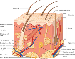
The skin is composed of two main layers: the epidermis and the dermis. The hypodermis (subcutaneous layer) lies beneath the dermis but is not technically part of the integumentary system.
Epidermis: Outermost, avascular layer made of keratinized stratified squamous epithelium.
Dermis: Vascular connective tissue layer housing hair follicles, glands, and sensory receptors.
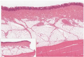
Hypodermis: Subcutaneous tissue composed of loose areolar and adipose tissue, connecting skin to underlying structures.
Epidermal Layers
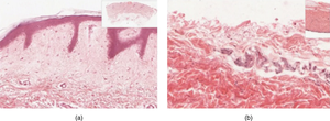
The epidermis is organized into distinct layers, each with specialized cells and functions. Thick skin (palms, soles) has five layers; thin skin has four.
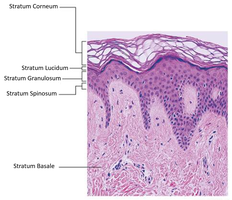
Stratum Basale: Deepest layer; single row of mitotically active basal cells, melanocytes (produce melanin), and Merkel cells (touch receptors).
Stratum Spinosum: 8-10 layers; contains keratinocytes and Langerhans cells (immune function).
Stratum Granulosum: 3-5 layers; cells produce keratin and keratohyalin, begin to die as organelles disintegrate.
Stratum Lucidum: Thin, clear layer found only in thick skin; contains eleidin protein.
Stratum Corneum: Outermost; 15-30 layers of dead, keratinized cells providing a tough, protective barrier.
Dermis and Hypodermis
Dermal Structure
The dermis is divided into two layers:
Papillary Layer: Superficial; loose areolar connective tissue, contains dermal papillae (form fingerprints), Meissner corpuscles (touch receptors), and capillaries.
Reticular Layer: Deep; dense irregular connective tissue, rich in collagen and elastin fibers, houses blood vessels, nerves, and glands.
The hypodermis is composed of loose areolar and adipose tissue, providing insulation, energy storage, and cushioning.
Accessory Structures
Hair
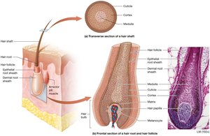
Hair is composed of dead, keratinized cells and originates from hair follicles in the dermis. It serves protective, sensory, and thermoregulatory functions.
Hair Shaft: Visible part above the skin surface.
Hair Root: Embedded in the follicle.
Hair Bulb: Base of the follicle; contains the hair matrix (mitotically active cells) and hair papilla (vascularized connective tissue).
Arrector Pili Muscle: Smooth muscle that causes hair to stand (goosebumps).
Nails
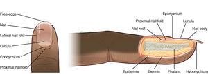
Nails are hard, keratinized structures that protect the distal tips of fingers and toes. They are formed by specialized keratinocytes in the nail matrix.
Nail Plate: Main body of the nail.
Nail Root: Proximal part under the skin; site of nail growth.
Nail Folds: Skin folds that anchor the nail.
Eponychium (Cuticle): Proximal nail fold.
Hyponychium: Area beneath the free edge of the nail.
Glands
The skin contains two main types of glands: sudoriferous (sweat) and sebaceous (oil) glands.
Eccrine Sweat Glands: Widely distributed; secrete watery sweat for thermoregulation.
Apocrine Sweat Glands: Found in axillae and genital regions; secrete protein-rich fluid into hair follicles, active after puberty.
Ceruminous Glands: Modified apocrine glands in the ear; produce cerumen (earwax).
Mammary Glands: Specialized sweat glands producing milk.
Sebaceous Glands: Secrete sebum (oil) to lubricate and waterproof skin and hair; have antimicrobial properties.
Functions of the Integumentary System
Protection and Sensation
The skin protects against environmental hazards and contains sensory receptors for touch, temperature, and pain. Meissner corpuscles detect light touch, while Pacinian corpuscles sense vibration.
Thermoregulation
The skin regulates body temperature through sweat production and control of blood flow to the skin surface. The sympathetic nervous system mediates these responses.
Sweating: Promotes heat loss via evaporation.
Vasodilation: Increases heat loss through radiation and convection.
Vasoconstriction: Conserves heat by reducing blood flow near the surface.
Vitamin D Synthesis
Exposure to UV radiation stimulates the skin to synthesize vitamin D, essential for calcium absorption and bone health.
Clinical Aspects
Skin Cancer
Skin cancer results from uncontrolled cell division, often due to UV exposure. There are three main types:
Basal Cell Carcinoma: Most common; arises from stratum basale.
Squamous Cell Carcinoma: Second most common; arises from stratum spinosum.
Melanoma: Most dangerous; arises from melanocytes, highly metastatic.
The ABCDE rule helps identify melanoma: Asymmetry, Border irregularity, Color variation, Diameter >6mm, Evolving shape.
Other Skin Disorders
Eczema: Allergic reaction causing dry, itchy, inflamed skin.
Acne Vulgaris: Inflammation of hair follicles due to excess sebum and keratin, often with bacterial involvement.
Burns and Injury
Burns are classified by depth:
First Degree: Epidermis only; redness, no scarring.
Second Degree: Epidermis and dermis; blistering, pain, possible scarring.
Third Degree: Full thickness (epidermis, dermis, hypodermis); severe damage.
Fourth Degree: Extends into muscle/bone.
The Rule of Nines divides the body into regions, each representing 9% of total body surface area, to estimate burn extent.
Other Skin Injuries
Pressure Injuries (Bedsores): Tissue necrosis from prolonged pressure.
Stretch Marks: Tearing of dermal collagen due to rapid stretching.
Callus: Thickened skin from increased friction.
Scar/Keloid: Collagen-rich tissue formed during wound healing; keloids are raised due to excess collagen.
