 Indietro
Lecture 6 - The Integumentary System: Structure, Function, and Clinical Aspects
Guida di studio - Note intelligenti
 Appunti personalizzati basati sui tuoi materiali, ampliati con definizioni chiave, esempi e contesto.
The Integumentary System
Overview of Skin
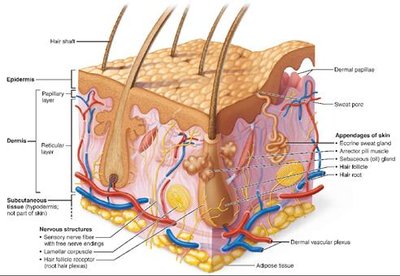
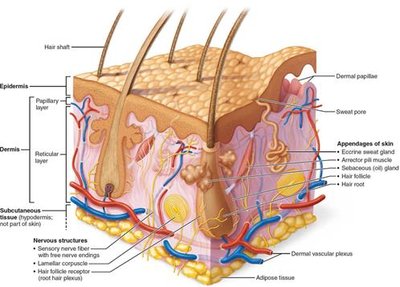
The skin is the largest organ of the human body and serves as a protective barrier between the internal environment and the external world. It consists of two main layers: the epidermis (outermost protective shield) and the dermis (underlying supportive layer). Beneath these lies the subcutaneous tissue (hypodermis or superficial fascia), which anchors the skin to underlying structures and provides insulation and cushioning.
Cells of the Epidermis
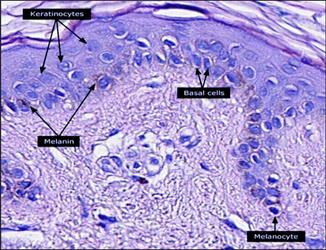
Major Cell Types
Keratinocytes: Produce keratin, a protein that gives skin its protective properties.
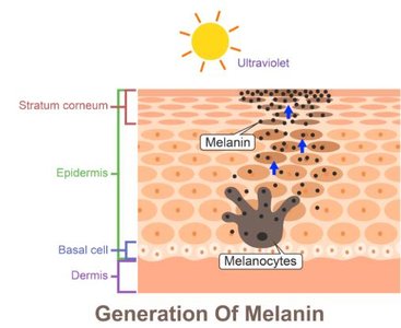
Melanocytes: Synthesize melanin, the pigment responsible for skin color and UV protection.
Dendritic (Langerhans) Cells: Immune cells that patrol the epidermis for pathogens.
Tactile Epithelial (Merkel) Cells: Sensory receptors for touch.
Layers of the Epidermis
Five Distinct Layers
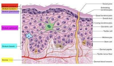
The epidermis is composed of five layers (from deep to superficial):
Stratum basale
Stratum spinosum
Stratum granulosum
Stratum lucidum (only in thick skin)
Stratum corneum
Stratum Basale
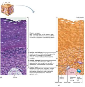
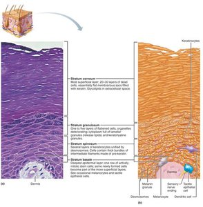
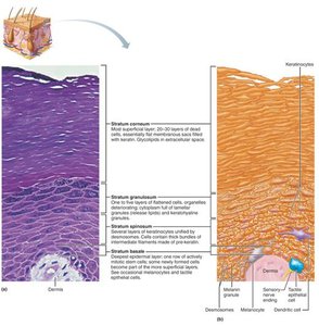
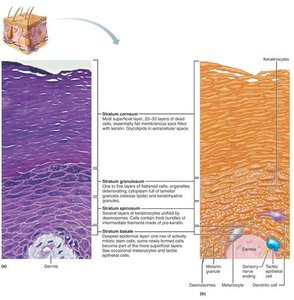
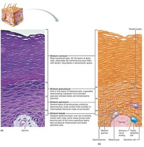
The deepest epidermal layer, firmly attached to the dermis, consists of a single row of stem cells that are highly mitotic, producing new keratinocytes. Melanocytes and tactile cells are also present.
Stratum Spinosum
This layer is several cells thick, with keratinocytes connected by desmosomes, giving a spiny appearance. It contains abundant melanosomes and dendritic cells, contributing to immune defense and pigment distribution.
Stratum Granulosum
Composed of 4–6 layers of flattened cells, this is where keratinization begins. Cells accumulate keratohyalin granules and lamellar granules, which help form a waterproof barrier. Cells above this layer die due to lack of nutrients.
Stratum Corneum
The outermost layer, consisting of 20–30 layers of dead, keratinized cells. These cells provide a durable overcoat for the body, protecting deeper layers from abrasion, pathogens, and water loss.
The Dermis
Structure and Function
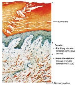
The dermis is a strong, flexible connective tissue layer beneath the epidermis. It contains blood vessels, nerves, hair follicles, and glands. The dermis is divided into two layers:
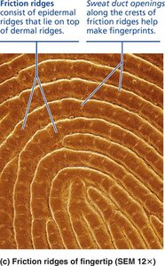
Papillary Dermis: Thin, superficial layer of areolar connective tissue with dermal papillae that project into the epidermis, forming friction ridges (fingerprints).
Reticular Dermis: Deeper, thicker layer of dense irregular connective tissue providing strength and elasticity.
Papillary Dermis and Friction Ridges
Dermal papillae increase the surface area for exchange of gases, nutrients, and waste products. In thick skin, friction ridges formed by dermal papillae enhance grip and tactile sensitivity.
Clinical Homeostatic Imbalances of the Skin
Stretch Marks and Blisters
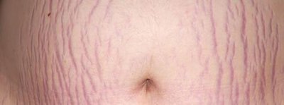
Striae: Silvery-white scars caused by dermal tearing due to extreme stretching (e.g., pregnancy, rapid weight gain).
Blisters: Fluid-filled pockets separating the epidermal and dermal layers, usually due to acute trauma.
Skin Color Determination
Melanin
Melanin is the only pigment produced in the skin, synthesized by melanocytes and transferred to keratinocytes. It protects DNA from UV damage. Melanin comes in two forms: reddish-yellow (pheomelanin) and brownish-black (eumelanin).
Carotene and Hemoglobin
Carotene: Yellow to orange pigment, most visible in the palms and soles, and can be converted to vitamin A.
Hemoglobin: Oxygen-carrying pigment in red blood cells; gives fair skin a pinkish hue due to low melanin levels.
Clinical Imbalances: Effects of Sun Exposure
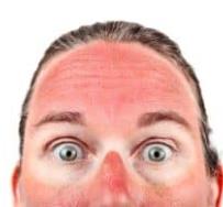
Excessive UV exposure can damage skin, cause DNA mutations (leading to cancer), destroy folic acid, and depress the immune system.
Photosensitivity is an abnormal reaction to sunlight.
Alterations in Skin Color as Clinical Indicators
Cyanosis (blue): Low oxygenation, indicating respiratory or cardiovascular issues.
Pallor (pale): Stress, anemia, or low blood pressure.
Erythema (red): Fever, inflammation, or embarrassment.
Jaundice (yellow): Liver disorders.
Bruises (ecchymoses/hematomas): Blood leakage under the skin.
Hyperpigmentation: May indicate endocrine disorders.
Hair
Structure and Function
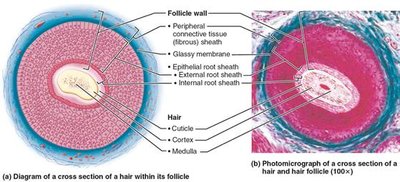
Hair is composed of dead, keratinized cells. It serves various functions, including sensory detection, protection from trauma, heat loss, and sunlight, as well as filtering particles (eyelashes, nose hairs).
Nails
Structure and Clinical Significance
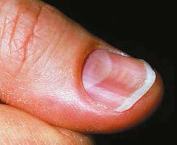
Nails are protective coverings on the dorsal surface of fingers and toes. Their appearance can indicate systemic diseases (e.g., yellow nails in respiratory disorders, spoon nails in iron deficiency, Beau's lines in severe illness).
Sweat Glands
Types and Functions
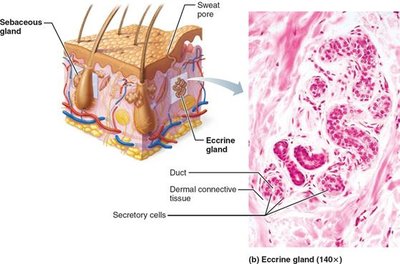
Eccrine Sweat Glands: Most numerous, found on palms, soles, and forehead. They secrete watery sweat for thermoregulation and excretion.
Apocrine Sweat Glands: Located in axillary and anogenital areas, secrete a viscous fluid into hair follicles; become active at puberty.
Sebaceous (Oil) Glands
Structure and Function
Sebaceous glands are widely distributed except in thick skin. They secrete sebum (an oily substance) into hair follicles, which lubricates and softens skin and hair and has bactericidal properties. Activity increases at puberty due to androgens.
Functions of the Skin
Major Roles
Protection: Chemical, physical, and biological barriers against pathogens, chemicals, and UV radiation.
Body Temperature Regulation: Through sweating and blood flow adjustments.
Cutaneous Sensations: Sensory receptors detect touch, temperature, pain, and pressure.
Metabolic Functions: Synthesis of vitamin D precursor, activation of steroid hormones.
Blood Reservoir: Stores up to 5% of body’s blood volume.
Excretion: Removal of nitrogenous wastes and salt via sweat.
Skin Cancer
Types and Risk Factors
Most skin tumors are benign, but some can metastasize. Major risk factors include UV exposure and chronic irritation. The three main types are:
Basal Cell Carcinoma: Most common, least malignant; arises from stratum basale.
Squamous Cell Carcinoma: Arises from keratinocytes of the stratum spinosum.
Melanoma: Most dangerous; arises from melanocytes.
Burns
Classification and Treatment
Burns are tissue injuries caused by heat, chemicals, electricity, or radiation. The main immediate threat is dehydration and electrolyte imbalance. The rule of nines is used to estimate the extent of burns and fluid loss. Burns are classified as critical if:
More than 25% of body has second-degree burns
More than 10% of body has third-degree burns
Face, hands, or feet have third-degree burns
Treatment includes debridement, antibiotics, temporary coverings, and skin grafts.
