 Indietro
The Integumentary System: Structure, Function, and Appendages
Guida di studio - Note intelligenti
 Appunti personalizzati basati sui tuoi materiali, ampliati con definizioni chiave, esempi e contesto.
The Integumentary System
Overview and Objectives
The integumentary system is composed of the skin and its appendages, serving as the body's primary interface with the external environment. This system is essential for protection, sensation, temperature regulation, and other vital functions. The following study notes cover the anatomy and functions of the skin, its appendages, pigmentation, and common homeostatic imbalances.
Functions of the Integumentary System
Key Functions
Protection: Acts as a chemical, physical, and mechanical barrier against pathogens, UV radiation, and physical injury.
Body Temperature Regulation: Achieved through dilation (cooling) and constriction (warming) of dermal vessels, and increased sweat gland activity.
Cutaneous Sensation: Exteroceptors in the skin detect touch, pain, and temperature changes.
Metabolic Functions: Synthesis of vitamin D occurs in the skin upon exposure to sunlight.
Blood Reservoir: Skin blood vessels can store up to 5% of the body's blood volume.
Excretion: Limited amounts of nitrogenous wastes are eliminated in sweat.
Anatomy of the Skin (Integument)
Structural Layers
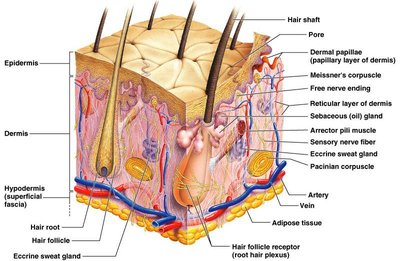
The skin consists of three primary layers: the epidermis, dermis, and hypodermis. Each layer has distinct structural and functional characteristics.
Epidermis
The epidermis is the outermost layer, exposed to the environment and responsible for protection. It is composed of keratinized stratified squamous epithelium, containing four distinct cell types and four or five layers.
Keratinocytes: Produce the fibrous protein keratin, providing strength and waterproofing.
Melanocytes: Produce melanin, the pigment responsible for skin color.
Langerhans' Cells: Epidermal macrophages that activate the immune system.
Merkel Cells: Function as touch receptors.
Layers of the Epidermis
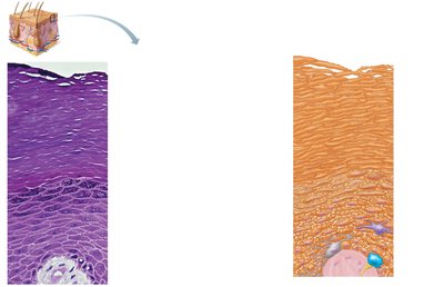
Stratum Corneum: Most superficial layer; consists of 20–30 layers of dead cells filled with keratin.
Stratum Granulosum: Three to five layers of flattened cells; contains lamellated and keratohyaline granules.
Stratum Spinosum: Several layers of keratinocytes unified by desmosomes; contains pre-keratin filaments.
Stratum Basale: Deepest layer; one row of actively mitotic stem cells, occasional melanocytes, and dendritic cells.
Stratum Lucidum: Present only in thick skin; clear layer of flattened dead keratinocytes.
Dermis
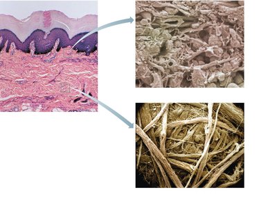
The dermis is the second major skin region, composed of strong, flexible connective tissue. It contains fibroblasts, macrophages, mast cells, and white blood cells. The dermis consists of two layers: papillary and reticular.
Papillary Layer: Areolar connective tissue with collagen and elastic fibers; contains dermal papillae, capillary loops, Meissner’s corpuscles, and free nerve endings.
Reticular Layer: Accounts for 80% of skin thickness; collagen fibers provide strength and resiliency, elastin fibers allow stretchability.
Hypodermis
The hypodermis (subcutaneous layer) lies deep to the skin and is composed of adipose and areolar connective tissue. It anchors the skin to underlying structures and provides insulation and cushioning.
Dermal Modifications and Skin Markings
Characteristic Skin Markings
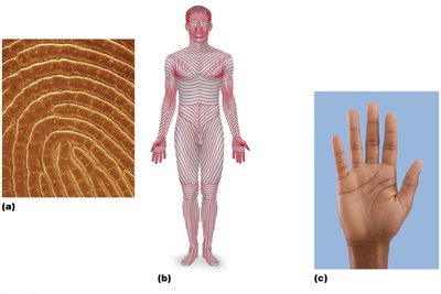
Friction Ridges: Enhance grip and are responsible for fingerprints.
Flexure Lines: Occur at joints where the skin is tightly secured to underlying structures.
Cleavage Lines: Indicate the direction of collagen fibers in the dermis, important for surgical incisions.
Appendages of the Skin
Cutaneous Glands
Skin appendages include sebaceous and sweat glands, which play roles in lubrication, temperature regulation, and excretion.
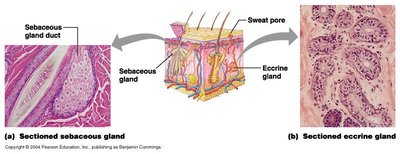
Sebaceous Glands: Simple alveolar glands found throughout the body; secrete sebum to soften skin and hair.
Sweat Glands (Sudoriferous): Three types:
Eccrine: Found in palms, soles, and forehead; involved in temperature regulation.
Apocrine: Located in axillary and anogenital areas; may act as scent glands.
Ceruminous: Modified apocrine glands in the ear; secrete cerumen (earwax).
Mammary Glands: Specialized sweat glands that secrete milk.
Summary Table: Cutaneous Glands
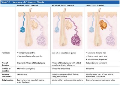
The following table summarizes the main types of cutaneous glands, their functions, methods of secretion, and locations.
Gland Type | Functions | Method of Secretion | Secretion Exits Duct At | Body Location |
|---|---|---|---|---|
Eccrine Sweat Glands | Temperature control, antibacterial properties | Merocrine (exocytosis) | Skin surface | Everywhere, especially palms, soles, forehead |
Apocrine Sweat Glands | May act as sexual scent glands | Merocrine (exocytosis) | Upper part of hair follicle; rarely skin surface | Mostly axillary and anogenital regions |
Sebaceous Glands | Lubricate skin and hair, prevent water loss, antibacterial properties | Holocrine | Upper part of hair follicle; sometimes skin surface | Everywhere except palms and soles |
Nails
Nails are scale-like modifications of the epidermis found on the distal, dorsal surfaces of fingers and toes. They protect the tips of digits and aid in picking up small objects.
Nail Matrix: Responsible for nail growth.
Body of Nail: Visible attached portion.
Free Edge: Distal portion that extends beyond the finger or toe.
Cuticle (Eponychium): Protective fold of skin at the base of the nail.

Hair
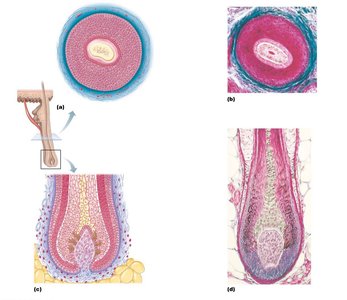
Hair consists of filamentous strands of dead keratinized cells produced by hair follicles. It contains hard keratin, which is tougher and more durable than the soft keratin of the skin. Hair is made up of the shaft (projecting from the skin) and the root (embedded in the skin).
Structure: Core (medulla), cortex, and cuticle.
Pigmentation: Provided by melanocytes at the base of the hair.
Functions: Maintains warmth, alerts the body to insects, guards the scalp against trauma, heat loss, and sunlight.
Distribution: Found everywhere except palms, soles, lips, nipples, and parts of external genitalia.
Types: Vellus (body hair), terminal (eyebrows and scalp).
Homeostatic Disturbances: Includes alopecia and male pattern baldness.

Pigmentation of Skin
Skin Color Determinants
Three pigments contribute to skin color:
Melanin: Ranges from yellow to reddish-brown to black; responsible for dark skin colors. Freckles and moles are local accumulations of melanin.
Carotene: Yellow to orange pigment, most obvious in the palms and soles.
Hemoglobin: Reddish pigment responsible for the pinkish hue of the skin.

Additional info:
Homeostatic imbalances such as skin cancers and burns are important clinical considerations, but detailed coverage is referenced for further reading (pp 165-167; Fig 5.11-13).
