 Indietro
The Integumentary System: Structure, Function, and Clinical Aspects
Guida di studio - Note intelligenti
 Appunti personalizzati basati sui tuoi materiali, ampliati con definizioni chiave, esempi e contesto.
The Integumentary System
Overview
The integumentary system consists of the skin and its derivatives, including sweat and oil glands, hair, and nails. This complex set of organs serves primarily protective functions and is essential for maintaining homeostasis.
Skin covers the entire body, with a surface area of 1.2 to 2.2 m2 and weighs 4 to 5 kg (about 7% of total body weight).
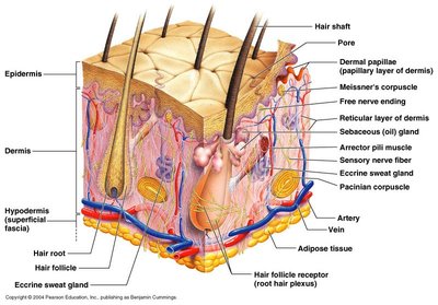
Three major regions: Epidermis (outermost), Dermis (middle), Hypodermis (deepest).
Structure of the Skin
Epidermis
The epidermis is the outermost layer, composed of keratinized stratified squamous epithelium. It contains four distinct cell types and four or five layers, depending on skin thickness.
Keratinocytes: Produce keratin, a fibrous protein for protection.
Melanocytes: Located in the deepest epidermis; produce melanin, which protects against UV radiation.
Langerhans’ cells: Epidermal macrophages involved in immune response.
Merkel cells: Touch receptors at the epidermal-dermal junction.
Layers of the Epidermis
Stratum basale (Basal Layer): Deepest layer, single row of young keratinocytes, rapid cell division, contains melanocytes and Merkel cells.
Stratum spinosum (Prickly Layer): Several cell layers thick, contains melanin granules and Langerhans’ cells, keratinocytes appear spiny.
Stratum granulosum (Granular Layer): Three to five cell layers, keratohyaline and lamellated granules accumulate, cells become more resistant.
Stratum lucidum (Clear Layer): Thin, transparent band, only in thick skin, few rows of flat, dead keratinocytes.
Stratum corneum (Horny Layer): Outermost layer, keratinized cells, waterproofing, protection from abrasion and penetration.
Dermis
The dermis is a strong, flexible connective tissue layer beneath the epidermis. It contains fibroblasts, macrophages, mast cells, and white blood cells.
Papillary layer: Areolar connective tissue, dermal papillae with capillary loops, Meissner’s corpuscles, and free nerve endings. Forms epidermal ridges (fingerprints).
Reticular layer: Dense irregular connective tissue, accounts for 80% of skin thickness, collagen fibers provide strength, elastin fibers allow stretch and recoil.
Hypodermis (Superficial Fascia)
The hypodermis is the subcutaneous layer deep to the skin, composed of adipose and areolar connective tissue. It anchors the skin to underlying structures and provides insulation and energy storage.
Skin Color
Pigments
Three pigments contribute to skin color:
Melanin: Yellow to reddish-brown to black pigment, responsible for dark skin colors. Freckles and moles are local accumulations.
Carotene: Yellow to orange pigment, most obvious in palms and soles.
Hemoglobin: Reddish pigment, gives pinkish hue to skin.
Clinical Significance
Alterations in skin color can indicate disease: erythema (redness), pallor (blanching), jaundice (yellow), bronzing, bruises (black and blue marks).
Skin Appendages
Sweat Glands (Sudoriferous Glands)
Sweat glands help regulate body temperature and excrete waste.
Eccrine glands: Found in palms, soles, forehead; secrete watery sweat with salts, vitamin C, antibodies, dermicidin, and waste.
Apocrine glands: Found in axillary and anogenital areas; secrete thicker sweat.
Ceruminous glands: Modified apocrine glands in ear canal; secrete cerumen (earwax).
Mammary glands: Specialized sweat glands; secrete milk.
Sebaceous Glands
Sebaceous glands are simple alveolar glands found throughout the body except palms and soles.
Secrete sebum, an oily substance that softens skin and hair.
Stimulated by hormones; larger on face, neck, upper chest.
Nails
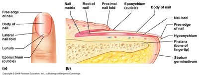
Nails are scalelike modifications of the epidermis on the distal, dorsal surface of fingers and toes.
Consist of nail matrix, root, body, free edge, and cuticle (eponychium).
Protect the tips of fingers and toes and aid in picking up small objects.
Hair
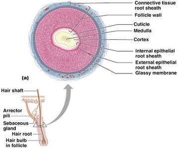
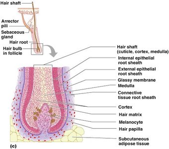
Hair consists of filamentous strands of dead keratinized cells produced by hair follicles.
Structure: Shaft (projects from skin), root (embedded in skin), medulla (core), cortex, cuticle (outermost).
Pigmentation: Melanocytes at base of hair.
Function: Maintains warmth, alerts to insects, guards scalp against trauma, heat loss, sunlight.
Distribution: All skin except palms, soles, lips, nipples, parts of external genitalia.
Types of Hair
Vellus: Pale, fine body hair found in children and adult females.
Terminal: Coarse, long hair of eyebrows, scalp, axillary, and pubic regions.
Hair Follicle
Root sheath extends from epidermal surface into dermis.
Deep end forms hair bulb; sensory nerve endings (root hair plexus) wrap around bulb.
Bending hair stimulates touch receptors.
Functions of the Integumentary System
Protection
The skin provides multiple layers of protection:
Chemical: Skin secretions (defensin, cathelicidins, melanin).
Physical: Keratinized cells form a mechanical barrier.
Biological: Langerhans cells, macrophages, and DNA.
Body Temperature Regulation
Dilation and constriction of dermal vessels regulate heat loss.
Increased sweat gland secretion cools the body.
Cutaneous Sensation
Exoreceptors sense touch and pain.
Metabolic Functions
Synthesis of vitamin D in dermal blood vessels.
Blood Reservoir
Skin blood vessels store up to 5% of body’s blood volume.
Excretion
Limited amounts of nitrogenous wastes eliminated in sweat.
Clinical Aspects
Homeostatic Imbalances
Acne: Inflammation of sebaceous glands, often due to bacterial infection.
Seborrhea: Overactive sebaceous glands, "cradle cap" in infants.
Hirsutism: Excessive hairiness due to androgen-secreting tumors.
Alopecia: Hair thinning in both sexes.
Male pattern baldness: Genetically determined, caused by follicular response to DHT.
Skin Cancer
Basal cell carcinoma: Least malignant, most common, arises from stratum basale.
Squamous cell carcinoma: Arises from keratinocytes of stratum spinosum, grows rapidly.
Melanoma: Cancer of melanocytes, highly metastatic, resistant to chemotherapy.
ABCD Rule for Melanoma
A: Asymmetry
B: Border irregularity
C: Color variation
D: Diameter > 6 mm
E: Elevation above skin surface
Burns
First-degree: Only epidermis damaged; redness, swelling, pain.
Second-degree: Epidermis and upper dermis damaged; blisters appear.
Third-degree: Entire skin thickness damaged; area appears gray-white, cherry red, or black; no initial pain.
Rule of Nines
Body divided into 11 areas (9% each) + 1% genital area.
Burns are critical if: >25% body has second-degree burns, >10% has third-degree burns, or third-degree burns on face, hands, feet.
Developmental Aspects of the Integument
Fetal Development
Epidermis: Develops from ectoderm.
Dermis and hypodermis: Develop from mesoderm.
Lanugo: Downy coat of delicate hairs covering fetus (5-6 months).
Vernix caseosa: Sebaceous gland secretion protecting fetal skin.
Adolescent to Adult
Skin and hair become oilier; acne may appear.
Environmental assaults accumulate; scaling and dermatitis more common.
Old Age
Epidermal cell replacement slows; skin thins.
Skin becomes dry, itchy; subcutaneous fat diminishes, leading to cold intolerance.
Decreased elasticity and subcutaneous tissue cause wrinkles.
Fewer melanocytes and Langerhans’ cells increase skin cancer risk.
