 Indietro
The Spinal Cord: Structure, Function, and Clinical Applications
Guida di studio - Note intelligenti
 Appunti personalizzati basati sui tuoi materiali, ampliati con definizioni chiave, esempi e contesto.
Central Nervous System: The Spinal Cord
Overview and Functions
The spinal cord is a critical component of the central nervous system, serving as a conduit for communication between the brain and the rest of the body. It also acts as a processing center for certain reflexes and local integration.
Relay station: Transmits sensory and motor information between the brain and peripheral nervous system.
Processing station: Integrates and processes some information locally, especially for spinal reflexes.
Protection of the Spinal Cord
Spinal Meninges and Spaces
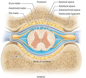
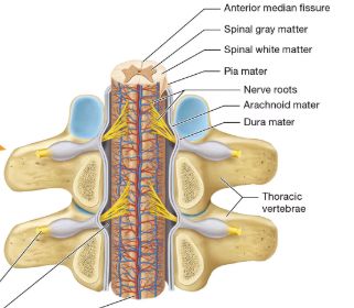
The spinal cord is protected by three connective tissue membranes called meninges, which are continuous with those of the brain but have some differences. These layers and the spaces between them are clinically significant.
Dura mater: In the spinal cord, only the meningeal layer is present (no periosteal layer as in the brain).
Arachnoid mater: Middle layer, same as in the brain.
Pia mater: Innermost layer, closely adheres to the spinal cord and forms denticulate ligaments that anchor the cord to the dura mater.
Space | Location | Contents/Clinical Use |
|---|---|---|
Epidural space | Between dura mater and vertebral canal | Fat, blood vessels; site for epidural anesthesia |
Subdural space | Between dura and arachnoid mater | Potential space (normally minimal fluid) |
Subarachnoid space | Between arachnoid and pia mater | Contains cerebrospinal fluid (CSF); site for lumbar puncture |
Clinical procedures:
Epidural anesthesia: Injection into the epidural space to block nerve transmission, commonly used in childbirth and surgeries.
Lumbar puncture (spinal tap): Needle inserted into the subarachnoid space (usually between L4 and L5) to collect CSF for diagnostic purposes.
Vertebral Column and Spinal Cord Anatomy
Regions of the Vertebral Column
The vertebral column is divided into regions, each with distinct vertebrae that articulate with the spinal cord and provide structural support.
Cervical vertebrae (C1–C7): Smallest vertebrae; C1 (Atlas) lacks a vertebral body and spinous process, C2 (Axis) has the dens, C7 has a prominent spinous process.
Thoracic vertebrae (T1–T12): Medium-sized bodies, long spinous processes, and facets for rib articulation.
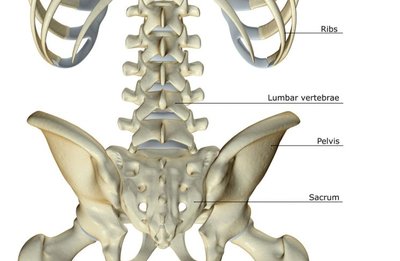
Lumbar vertebrae (L1–L5): Largest vertebral bodies, designed to support the most weight.
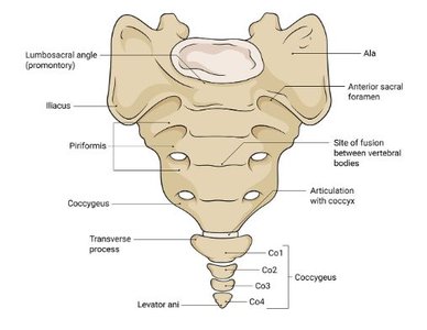
Sacrum: Fused vertebrae forming the posterior wall of the pelvis.
Coccyx: Small, fused bones at the base of the vertebral column (tailbone).
Vertebral Structure and Special Features
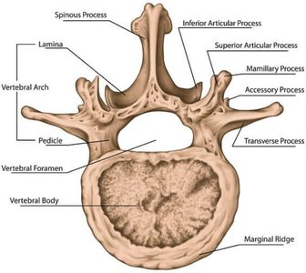
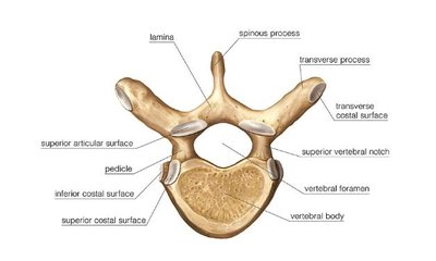
Each vertebra has characteristic features that allow for protection and passage of the spinal cord.
Vertebral body: Weight-bearing portion.
Vertebral foramen: Canal through which the spinal cord passes.
Spinous and transverse processes: Sites for muscle and ligament attachment.
Articular processes: Form joints with adjacent vertebrae.
Special Cervical Vertebrae
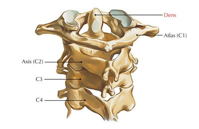
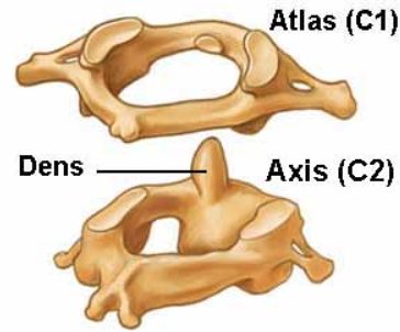
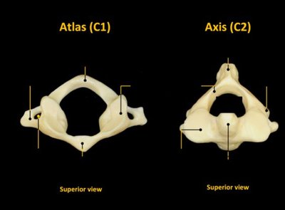
The first two cervical vertebrae, the atlas and axis, have unique structures to support the skull and allow head rotation.
Atlas (C1): Lacks a vertebral body and spinous process; supports the skull.
Axis (C2): Contains the dens (odontoid process), which acts as a pivot for rotation of the atlas and skull.
C7: Has a prominent spinous process (vertebra prominens).
External Anatomy of the Spinal Cord
Major Features and Extensions
The spinal cord extends from the foramen magnum to the level of L1–L2, where it tapers into the conus medullaris. Nerve roots continue as the cauda equina and the filum terminale anchors the cord to the coccyx.
Cervical and lumbar enlargements: Regions where nerves for the limbs arise.
Anterior median fissure: Deep groove on the anterior surface.
Posterior median sulcus: Shallow groove on the posterior surface.
Spinal nerves: 31 pairs, part of the peripheral nervous system, carry sensory and motor information.
Internal Anatomy of the Spinal Cord
Gray Matter and White Matter Organization
The spinal cord's internal structure is organized into gray and white matter, each with distinct regions and functions.
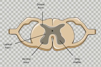
Central canal: Runs through the center, filled with CSF.
Gray commissure: Connects the two sides of gray matter.
Gray matter: Butterfly-shaped region containing neuron cell bodies.
White matter: Surrounds gray matter, contains myelinated axons organized into tracts.
Gray Matter Region | Function |
|---|---|
Anterior (ventral) horn | Somatic motor neurons (skeletal muscle control) |
Posterior (dorsal) horn | Sensory neurons (somatic and visceral sensory input) |
Lateral horn | Autonomic motor neurons (visceral motor control, only in thoracic and upper lumbar regions) |
White Matter: Funiculi and Tracts
White matter is divided into three regions called funiculi, each containing specific ascending (sensory) and descending (motor) tracts.
Posterior funiculus
Lateral funiculus
Anterior funiculus
Each funiculus contains multiple tracts (columns), which are bilateral and mirror each other.
Ascending (Sensory) Tracts
Major Sensory Pathways
Ascending tracts carry sensory information from the body to the brain. The main tracts include:
Ascending Tract | Function |
|---|---|
Posterior columns (fasciculus gracilis & cuneatus) | Somatosensory stimuli (fine touch, vibration, proprioception) |
Spinocerebellar tracts | Proprioceptive information to the cerebellum |
Anterolateral system (spinothalamic tracts) | Pain, temperature, and some touch stimuli |
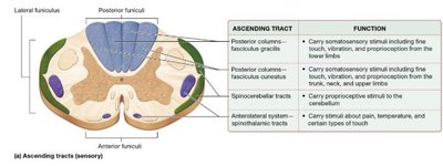
Example: The spinothalamic tract is responsible for transmitting pain and temperature sensations to the brain.
Descending (Motor) Tracts
Major Motor Pathways
Descending tracts carry motor commands from the brain to the body. The main tracts include:
Descending Tract | Function |
|---|---|
Corticospinal tracts | Voluntary control of skeletal muscles below the head and neck |
Reticulospinal tracts | Posture and limb orientation during movement |
Tectospinal tracts | Reflexive movements of the head and eyes |
Vestibulospinal tracts | Posture and balance |
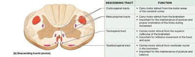
Example: The corticospinal tract originates in the cerebral cortex and is the primary pathway for voluntary motor control.
Clinical Applications
Epidural Anesthesia and Lumbar Puncture
Epidural anesthesia: Local anesthetic injected into the epidural space, causing numbness below the injection site by blocking sensory and motor nerve transmission.
Lumbar puncture: Needle inserted into the subarachnoid space (usually between L4 and L5) to collect CSF for diagnostic testing (e.g., meningitis, multiple sclerosis).
Practice Application
Question: Which sensory tract carries stimuli of pain, temperature, and some kinds of touch?
Answer: The anterolateral system (spinothalamic tracts) carries these stimuli. It is an ascending tract.
