 Indietro
Tissue: The Living Fabric – Study Notes for Anatomy & Physiology
Guida di studio - Note intelligenti
 Appunti personalizzati basati sui tuoi materiali, ampliati con definizioni chiave, esempi e contesto.
Tissues: The Living Fabric
Overview of Tissues
Tissues are groups of cells similar in structure and function, forming the basic fabric of the human body. There are four major types: epithelial (covering), connective (support), muscle (movement), and nervous (control). Each tissue type is specialized to help maintain homeostasis.
Histology: The study of tissues, complements gross anatomy, and is essential for understanding organ physiology.
Complementarity: The structure of tissues predicts their function.
Major Tissue Types and Functions
Nervous tissue: Internal communication (brain, spinal cord, nerves).
Muscle tissue: Contracts to cause movement (skeletal, cardiac, smooth muscle).
Epithelial tissue: Forms boundaries, protects, secretes, absorbs, filters, excretes (skin, GI tract lining).
Connective tissue: Supports, protects, binds other tissues together (bones, tendons, fat).
Epithelial Tissue
Definition and Types
Epithelial tissue (epithelium) is a sheet of cells covering body surfaces or cavities. It is classified by location:
Covering and lining epithelium: Forms outer skin and lines body cavities.
Glandular epithelium: Secretory tissue in glands.
Functions of Epithelial Tissue
Protection (skin)
Absorption (digestive tract)
Filtration (kidneys)
Excretion (kidneys)
Secretion (glands)
Sensory reception (temperature, pressure)
Characteristics of Epithelial Tissue
Polarity: Apical (free) and basal (attached) surfaces. Apical surfaces may have microvilli or cilia.
Basal lamina: Non-cellular layer adjacent to basal surface, supports and acts as a selective filter.
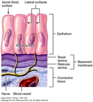
Cellularity: Composed of closely packed cells, held together by tight junctions and desmosomes.
Supported by connective tissue: Reticular lamina under basal lamina; together form the basement membrane.
Avascular but innervated: No blood vessels, but has nerve supply; receives nutrients by diffusion.
High rate of regeneration: Rapidly replaces lost or damaged cells.
Classification of Epithelia
Epithelia are classified by number of layers and cell shape:
Layers: Simple (one layer), Stratified (multiple layers)
Shape: Squamous (flat), Cuboidal (cube-shaped), Columnar (tall)
If stratified, named according to the apical layer of cells.
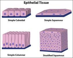
Types of Epithelial Tissue
Simple Squamous Epithelium: Single layer of flattened cells; allows diffusion and filtration; found in kidney glomeruli, air sacs of lungs, lining of heart, blood vessels, and serosae.
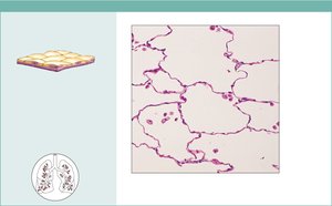
Simple Cuboidal Epithelium: Single layer of cube-shaped cells; functions in secretion and absorption; found in small ducts and kidney tubules.
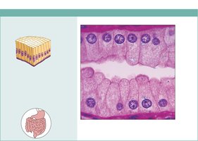
Simple Columnar Epithelium: Single layer of tall cells; absorption and secretion; ciliated type propels mucus; found in digestive tract, gallbladder, small bronchi, uterine tubes.
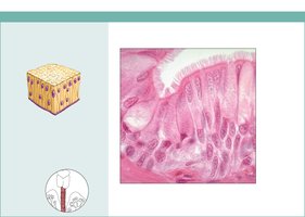
Pseudostratified Columnar Epithelium: Single layer of cells of varying heights; nuclei at different levels; secretion and propulsion of mucus; ciliated type lines trachea and upper respiratory tract.
Stratified Squamous Epithelium: Several layers; protects against abrasion; nonkeratinized type lines esophagus, mouth, vagina; keratinized type forms epidermis of skin.
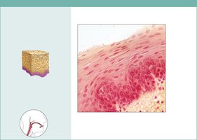
Stratified Cuboidal Epithelium: Rare; found in sweat and mammary glands.
Stratified Columnar Epithelium: Limited distribution; found in pharynx, male urethra, glandular ducts.
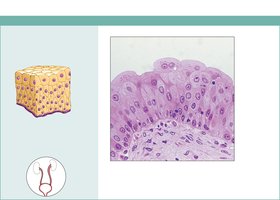
Transitional Epithelium: Resembles both stratified squamous and cuboidal; stretches and permits distension; lines ureters, bladder, part of urethra.
Glandular Epithelia
A gland is one or more cells that makes and secretes an aqueous fluid. Classified by:
Site of product release: Endocrine (ductless, hormones) or exocrine (with ducts, e.g., sweat, oil).
Number of cells: Unicellular (e.g., goblet cells) or multicellular.
Modes of Secretion
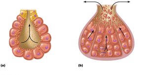
Merocrine glands: Secrete products by exocytosis (e.g., pancreas, sweat glands).
Holocrine glands: Accumulate products until cell ruptures (e.g., sebaceous glands).
Connective Tissue
Overview and Classes
Connective tissue is the most abundant and widely distributed tissue type. It supports, protects, and binds other tissues. Four main classes:
Connective tissue proper (dense & loose)
Cartilage
Bone tissue
Blood
Major Functions
Binding and support
Protection
Insulation
Transportation (blood)
Storing reserve fuel (fat)
Characteristics of Connective Tissue
All originate from mesenchyme (embryonic tissue).
Varying degrees of vascularity.
Composed largely of non-living extracellular matrix (ground substance and fibers).
Structural Elements
Ground substance: Material between cells, allows diffusion of nutrients.
Fibers: Collagen (strength), elastic (stretch), reticular (support).
Cells: "Blast" cells (immature, matrix-producing), "cyte" cells (mature, maintain matrix), adipocytes (store nutrients), white blood cells, mast cells, macrophages.
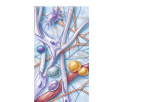
Connective Tissue Proper
Loose Connective Tissue
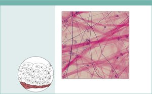
Areolar: Gel-like matrix with all three fibers; wraps and cushions organs; widely distributed under epithelia.
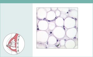
Adipose: Closely packed fat cells; stores energy, insulates, supports organs; found under skin, around kidneys, in breasts.
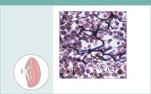
Reticular: Network of reticular fibers; forms internal skeleton for lymphoid organs.
Dense Connective Tissue
Dense regular: Parallel collagen fibers; attaches muscles to bones; found in tendons, ligaments.
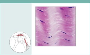
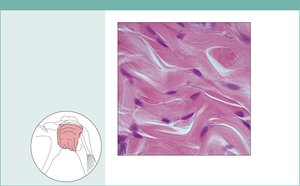
Dense irregular: Irregularly arranged collagen fibers; withstands tension in many directions; found in dermis, joint capsules.
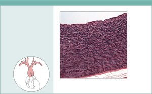
Elastic: High proportion of elastic fibers; allows recoil; found in walls of arteries, bronchial tubes.
Cartilage
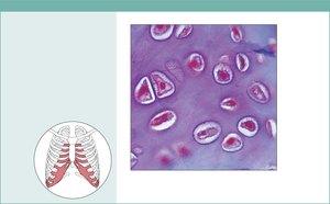
Hyaline cartilage: Most abundant; supports, cushions; found at ends of long bones, nose, trachea.
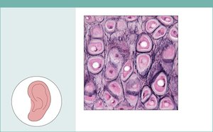
Elastic cartilage: More elastic fibers; maintains shape, flexibility; found in ear, epiglottis.
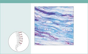
Fibrocartilage: Thick collagen fibers; strong, absorbs shock; found in intervertebral discs, knee.
Bone (Osseous Tissue)
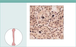
Hard, calcified matrix; supports and protects; stores fat and minerals; site of blood cell formation.
Osteoblasts produce matrix; osteocytes maintain it; reside in lacunae.
Blood
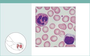
Fluid matrix (plasma); transports gases, nutrients, wastes.
Contains red and white blood cells.
Nervous Tissue
Structure and Function
Nervous tissue is the main component of the nervous system (brain, spinal cord, nerves). It regulates and controls body functions. Two main cell types:
Neurons: Generate and conduct nerve impulses; consist of cell body, axon, dendrites.
Glial cells: Support, insulate, and protect neurons.
Muscle Tissue
Types and Functions
Muscle tissue is highly vascularized and responsible for movement. Muscle cells contain myofilaments (actin and myosin) for contraction. Three types:
Skeletal muscle: Long, cylindrical, multinucleate, striated; voluntary movement.
Cardiac muscle: Branching, striated, uninucleate; involuntary; intercalated discs.
Smooth muscle: Spindle-shaped, central nuclei, no striations; involuntary.
Membranes That Cover & Line
Types of Membranes
Cutaneous membrane: Skin; keratinized stratified squamous epithelium attached to dense irregular connective tissue.
Mucous membrane: Lines body cavities open to exterior; moist; stratified squamous or simple columnar epithelium with loose connective tissue.
Serous membrane: Double layer (parietal/visceral); lines closed ventral body cavities; simple squamous epithelium on areolar tissue; secretes serous fluid.
Tissue Repair
Steps in Tissue Repair
Inflammation: Release of chemicals, dilation of blood vessels, increased permeability, clotting, scab formation.
Organization & restored blood supply: Blood clot replaced with granulation tissue, epithelium regenerates, fibroblasts produce collagen, debris is phagocytized.
Regeneration and fibrosis: Surface epithelium regenerates, scab detaches, scar tissue forms, epithelium thickens.
Regenerative Capacity of Tissues
High regeneration: Epithelium, mucosae, bone, areolar, dense irregular connective tissue, blood-forming tissue.
Moderate regeneration: Smooth muscle, dense regular connective tissue.
Weak regeneration: Skeletal muscle, cartilage.
No regeneration: Cardiac muscle, nervous tissue in brain and spinal cord.
Developmental Biology
Epithelium arises from all three germ layers.
Nervous tissue from ectoderm.
Muscle and connective tissue from mesoderm.
Clinical Homeostatic Imbalance
Scar tissue in organs can impair function, block substances, interfere with muscle contraction or nerve transmission.
Scar adhesions may cause organs to adhere to neighboring structures, potentially causing progressive organ failure.
