 Indietro
Tissues: The Living Fabric – Structure and Function of Human Tissues
Guida di studio - Note intelligenti
 Appunti personalizzati basati sui tuoi materiali, ampliati con definizioni chiave, esempi e contesto.
Tissues: The Living Fabric
Introduction to Tissues and Histology
Tissues are groups of cells similar in structure that perform common or related functions. The study of tissues and their cellular organization is known as histology. Each tissue type is specialized to carry out specific roles essential for maintaining life in multicellular organisms.
Tissue: Groups of similar cells performing a common function.
Histology: The microscopic study of tissue structure and organization.
Specialization: Individual body cells are specialized for particular functions.
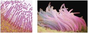
Primary Tissue Types
Overview of the Four Basic Tissue Types
The human body is composed of four primary tissue types, each with distinct structures and functions:
Epithelial Tissue: Covers body surfaces, lines cavities, and forms glands.
Connective Tissue: Supports, protects, binds other tissues, stores energy, and transports substances.
Muscle Tissue: Contracts to produce movement.
Nervous Tissue: Initiates and transmits electrical impulses for communication.

Nervous Tissue
Structure and Function
Nervous tissue is the main component of the nervous system, including the brain, spinal cord, and nerves. It is specialized for the regulation and control of body functions through the generation and conduction of nerve impulses.
Neurons: Specialized cells that generate and conduct electrical impulses.
Neuroglia: Supporting cells that insulate, protect, and support neurons.

Muscle Tissue
Types and Functions
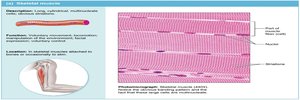
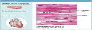
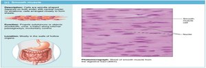
Muscle tissue is highly vascularized and responsible for most types of body movement. There are three types of muscle tissue, each with unique structural and functional characteristics:
Skeletal Muscle: Voluntary, striated muscle attached to bones for movement.
Cardiac Muscle: Involuntary, striated muscle found only in the heart, responsible for pumping blood.
Smooth Muscle: Involuntary, non-striated muscle found in the walls of hollow organs, responsible for propelling substances.
Epithelial Tissue (Epithelium)
Functions and Characteristics
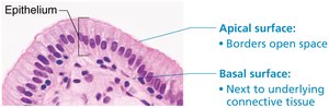
Epithelial tissue forms boundaries between different environments and serves several key functions, including protection, absorption, filtration, excretion, secretion, and sensory reception. It is classified based on location as covering/lining epithelia or glandular epithelia.
Polarity: Has an apical (free) surface and a basal (attached) surface.
Supported by Connective Tissue: The basal lamina anchors epithelium to underlying tissues.
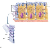
Specialized Contacts: Tight junctions and desmosomes bind adjacent cells.
Avascular but Innervated: Contains no blood vessels but is supplied by nerves.
Regeneration: High capacity for renewal due to frequent cell loss.


Classification of Epithelia
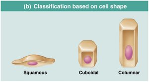
Epithelia are classified by the number of cell layers and the shape of the cells in the apical layer:
Simple Epithelium: Single layer of cells.
Stratified Epithelium: Two or more layers of cells.
Squamous Cells: Flattened and scale-like.
Cuboidal Cells: Box-like, as tall as they are wide.
Columnar Cells: Tall and column-shaped.
Types of Epithelial Tissue
Simple Squamous Epithelium: Single layer of flat cells; allows diffusion and filtration. Found in air sacs of lungs and lining of blood vessels.
Simple Cuboidal Epithelium: Single layer of cube-shaped cells; functions in secretion and absorption. Found in kidney tubules and glands.
Simple Columnar Epithelium: Single layer of tall cells; absorption and secretion. Lines most of the digestive tract.
Pseudostratified Columnar Epithelium: Appears stratified but is a single layer; secretion and propulsion of mucus. Found in trachea and upper respiratory tract.
Stratified Squamous Epithelium: Multiple layers; protects underlying tissues. Found in skin, mouth, and esophagus.
Transitional Epithelium: Resembles both stratified squamous and cuboidal; stretches readily. Lines urinary bladder and ureters.






Glandular Epithelium
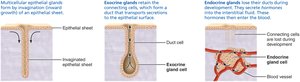
Glandular epithelium consists of cells specialized to produce and secrete substances. Glands are classified as exocrine (with ducts, secrete non-hormonal substances) or endocrine (ductless, secrete hormones).
Exocrine Glands: Secrete products into ducts (e.g., sweat, salivary glands).
Endocrine Glands: Secrete hormones directly into the bloodstream.

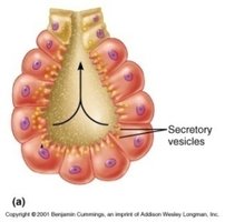
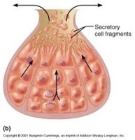
Modes of Secretion
Merocrine: Secretion by exocytosis (e.g., sweat glands).
Apocrine: Apex of cell pinches off (e.g., mammary glands).
Holocrine: Entire cell ruptures to release product (e.g., sebaceous glands).
Connective Tissue (CT)
Functions and Characteristics
Connective tissue is the most abundant and widely distributed tissue type in the body. It supports, binds, protects, insulates, stores energy, and transports substances. CT is unique in having an extensive extracellular matrix, which allows it to bear weight, withstand tension, and endure physical stress.
Common Origin: All CT arises from mesenchyme (embryonic tissue).
Vascularity: Varies from avascular (cartilage) to highly vascular (bone).
Extracellular Matrix: Composed of ground substance, fibers, and cells.


Structural Elements of Connective Tissue
Ground Substance: Unstructured material that fills the space between cells.
Fibers: Collagen (strength), elastic (stretch), and reticular (support).
Cells: Fibroblasts, chondroblasts, osteoblasts, adipocytes, and blood cells.

Classification of Connective Tissue
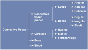
Connective tissue is classified into connective tissue proper, cartilage, bone, and blood. Each class has unique cells, fibers, and functions.

Tissue Class | Subclasses | Cells | Matrix | General Features |
|---|---|---|---|---|
Connective Tissue Proper | Loose (areolar, adipose, reticular); Dense (regular, irregular, elastic) | Fibroblasts, adipocytes | Gel-like ground substance, collagen, reticular, elastic fibers | Binding, support, energy storage |
Cartilage | Hyaline, elastic, fibrocartilage | Chondroblasts, chondrocytes | Gel-like ground substance, collagen, elastic fibers | Resists compression, supports body structures |
Bone | Compact, spongy | Osteoblasts, osteocytes | Hard, calcified matrix, collagen fibers | Support, protection, blood cell formation |
Blood | --- | Red and white blood cells, platelets | Plasma (fluid matrix) | Transport of gases, nutrients, wastes |

Connective Tissue Proper
Loose Connective Tissue: Areolar (packing material), adipose (fat storage), reticular (internal framework for lymphoid organs).
Dense Connective Tissue: Regular (tendons, ligaments), irregular (dermis), elastic (artery walls).

Cartilage
Cartilage is tough yet flexible, avascular, and lacks nerve fibers. It contains chondrocytes in lacunae and is surrounded by perichondrium. Types include hyaline, elastic, and fibrocartilage.
Hyaline Cartilage: Most abundant, supports and reinforces.
Elastic Cartilage: Maintains shape with flexibility.
Fibrocartilage: Absorbs compressive shock.
Bone (Osseous Tissue)
Bone supports and protects body structures, stores calcium and fat, and synthesizes blood cells. It has a calcified matrix with more collagen than cartilage and is richly vascularized.
Osteoblasts: Produce bone matrix.
Osteocytes: Maintain bone matrix.
Blood
Blood is an atypical connective tissue with a fluid matrix (plasma). It contains red blood cells (RBCs), white blood cells (WBCs), and platelets. Blood functions as a transport vehicle for nutrients, gases, wastes, and other substances throughout the body.
Developmental Origin of Tissues
Germ Layers and Tissue Differentiation
All tissues originate from three primary germ layers in the embryo: ectoderm, mesoderm, and endoderm. Epithelial tissue arises from all three layers, while connective and muscle tissues derive mainly from mesoderm, and nervous tissue from ectoderm.
Additional info: This guide expands on the provided slides with definitions, examples, and academic context to ensure completeness and clarity for Anatomy & Physiology students.
