Ch. 6 The Muscular System
Marieb - Essentials of Human Anatomy & Physiology 13th Edition
Marieb13th EditionEssentials of Human Anatomy & PhysiologyISBN: 9780135624340Non è quello che usi tu?Cambia libro di testo
Capitolo 6, Problema 1

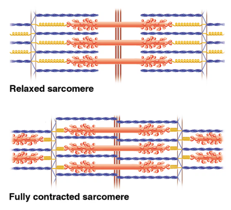
Compare these images of a relaxed skeletal muscle fiber and a fully contracted muscle fiber. Which would you see only in the relaxed fiber?
Diagram showing structural differences between relaxed and fully contracted sarcomeres in skeletal muscle fibers.
a. Z discs
b. Sarcomeres
c. I bands
d. A bands
e. H zones

Guida verificata passo dopo passo
1
Step 1: Identify the structural components in the relaxed sarcomere image. Notice the Z discs (dark vertical lines), I bands (light regions with thin filaments only), A bands (dark regions with thick filaments), and the H zone (lighter central region within the A band where there are only thick filaments).
Step 2: Compare these components to the fully contracted sarcomere image. Observe that the Z discs move closer together, the I bands and H zones become narrower or disappear, while the A bands remain the same length.
Step 3: Understand that the H zone is the region in the center of the A band where only thick filaments are present and no thin filaments overlap. During contraction, thin filaments slide over thick filaments, reducing or eliminating the H zone.
Step 4: Recognize that the I bands, which contain only thin filaments, also shorten during contraction as the thin filaments slide inward.
Step 5: Conclude that the H zone is visible only in the relaxed sarcomere and disappears in the fully contracted sarcomere, making option (e) H zones the correct answer.

Risposta video verificata per un problema simile:

Questa soluzione video è stata consigliata dai nostri tutor come utile per risolvere questo problema.
Durata del video:
1m

Concetti chiave

Ecco i concetti essenziali che devi comprendere per rispondere correttamente alla domanda.

Sarcomere Structure

The sarcomere is the basic contractile unit of a skeletal muscle fiber, defined by the area between two Z discs. It contains thick filaments (myosin) and thin filaments (actin), organized into distinct bands and zones that change during muscle contraction.
Video consigliato:
7:18
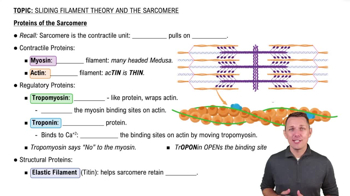
Proteins of the Sarcomere

Muscle Contraction and Sliding Filament Theory

Muscle contraction occurs when thin actin filaments slide over thick myosin filaments, shortening the sarcomere. This sliding reduces the width of the I bands and H zones, while the A band remains constant, reflecting the overlap of filaments.
Video consigliato:
6:26
Sliding Filament Theory

Identification of Sarcomere Bands and Zones

The Z discs mark sarcomere boundaries; I bands contain only thin filaments; A bands contain thick filaments and overlapping thin filaments; H zones are central regions of thick filaments without overlap. The H zone disappears during full contraction.
Video consigliato:
6:01
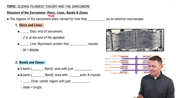
Structure of the Sarcomere: Bands, Zones, Discs & Lines