 Indietro
Introduction to Proteins: Structure, Properties, and Purification
Guida di studio - Note intelligenti
 Appunti personalizzati basati sui tuoi materiali, ampliati con definizioni chiave, esempi e contesto.
Introduction to Proteins
Overview of Protein Biosynthesis
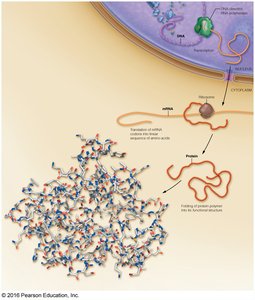
Proteins are essential macromolecules synthesized through a highly regulated process involving transcription and translation. The genetic information encoded in DNA is transcribed into messenger RNA (mRNA) in the nucleus. The mRNA is then exported to the cytoplasm, where ribosomes translate the nucleotide sequence into a linear chain of amino acids, which subsequently folds into a functional three-dimensional protein structure.
Amino Acids: Structure and Properties
General Structure of Amino Acids
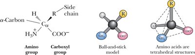
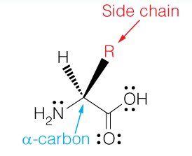
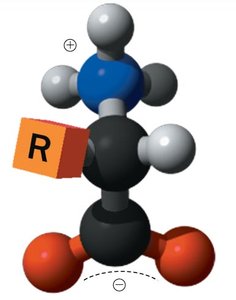
Amino acids are the building blocks of proteins. Each amino acid contains a central (α) carbon atom bonded to an amino group, a carboxyl group, a hydrogen atom, and a variable side chain (R group) that determines its unique properties. Except for glycine, all amino acids are chiral and exist as L- or D-enantiomers, with L-amino acids predominating in nature.
Zwitterionic Nature of Amino Acids
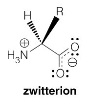
At physiological pH (~7), amino acids exist as zwitterions, where the amino group is protonated (NH3+) and the carboxyl group is deprotonated (COO-), resulting in a molecule with both positive and negative charges but an overall neutral charge.
Stereochemistry of Amino Acids
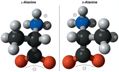
All amino acids except glycine are chiral, possessing an asymmetric α-carbon. The L- and D-forms are mirror images (enantiomers), but only L-amino acids are incorporated into proteins. The (R,S) system provides unambiguous nomenclature for amino acids with multiple chiral centers, such as isoleucine and threonine.
Classification of Amino Acids
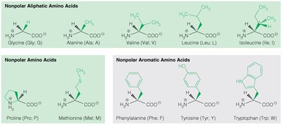
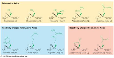
Amino acids are classified based on the chemical properties of their side chains:
Nonpolar aliphatic: Glycine, Alanine, Valine, Leucine, Isoleucine, Proline, Methionine
Nonpolar aromatic: Phenylalanine, Tyrosine, Tryptophan
Polar uncharged: Serine, Threonine, Cysteine, Asparagine, Glutamine
Positively charged (basic): Lysine, Arginine, Histidine
Negatively charged (acidic): Aspartic acid, Glutamic acid
Special Properties of Selected Amino Acids
Glycine: Smallest, non-chiral amino acid.
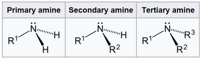
Proline: Contains a secondary amine, forms a rigid ring structure.
Serine, Threonine, Tyrosine: Side chains can be phosphorylated.
Cysteine: Contains a thiol group, can form disulfide bonds upon oxidation.
Histidine: Side chain pKa near physiological pH, important in enzyme catalysis.
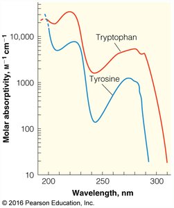
Tryptophan and Tyrosine: Major contributors to protein UV absorbance at 280 nm.
UV Absorbance of Aromatic Amino Acids
Tryptophan and tyrosine absorb UV light strongly at 280 nm, which is used to quantify protein concentration. Phenylalanine absorbs weakly and is less useful for this purpose. The ratio of absorbance at 260/280 nm helps distinguish proteins from nucleic acids.
Ionizable Side Chains and pKa Values
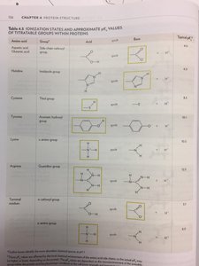
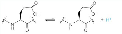
Some amino acids have ionizable side chains, which can gain or lose protons depending on the pH. The pKa value is the pH at which half of the group is ionized. The charge state of these side chains is crucial for protein structure and function.
Sidechain of Amino Acid | Charge in Acid Form | Charge in Basic Form | pKa |
|---|---|---|---|
Arg (R) | +1 | 0 | 12.5 |
Lys (K) | +1 | 0 | 10.0 |
His (H) | +1 | 0 | 6.0 |
Asp (D) | 0 | -1 | 3.9 |
Glu (E) | 0 | -1 | 4.2 |
Cys (C) | 0 | -1 | 8.3 |
Tyr (Y) | 0 | -1 | 10.1 |
Amino End | +1 | 0 | 9.0 |
Carboxylic End | 0 | -1 | 2.0 |
Titration Curves and Isoelectric Point (pI)
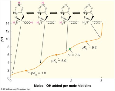
The titration curve of an amino acid or peptide shows how its net charge changes with pH. The isoelectric point (pI) is the pH at which the molecule carries no net charge. At pH values below the pI, the molecule is positively charged; above the pI, it is negatively charged.
Peptide Bond and Protein Primary Structure
Formation and Properties of the Peptide Bond
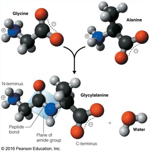
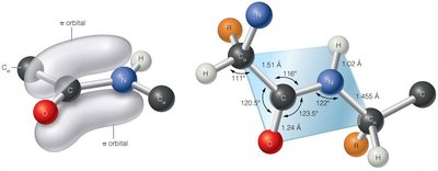
A peptide bond forms between the carboxyl group of one amino acid and the amino group of another, releasing water in a condensation reaction. This bond is planar and exhibits partial double-bond character due to resonance, making it non-rotatable and contributing to protein stability.
Peptide Sequence and Directionality
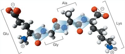
Peptide and protein sequences are always written from the amino (N-) terminus to the carboxyl (C-) terminus. Oligopeptides contain a few residues, while polypeptides have more than 15–20 residues. Sequence determines the unique properties and function of each protein.
Calculation of Peptide Charge
Determining Net Charge at a Given pH
To calculate the net charge of a peptide at a specific pH:
Identify all ionizable groups (side chains, N-terminus, C-terminus).
Compare the pH to the pKa of each group to determine its charge state.
Sum the charges of all ionizable groups to obtain the net charge.
Protein Purification Strategies
Affinity Chromatography
Affinity chromatography exploits specific interactions between a protein and a ligand attached to a stationary phase. For example, His-tagged proteins bind to Ni2+ or Zn2+ columns and are eluted with imidazole.
Ion Exchange Chromatography (IEC)
IEC separates proteins based on their net surface charge. Cation exchangers bind positively charged proteins, while anion exchangers bind negatively charged proteins. Proteins are eluted by increasing salt concentration or changing pH.
Size Exclusion Chromatography (SEC)
SEC separates proteins based on size. Larger proteins elute first because they are excluded from the pores of the matrix, while smaller proteins are retained longer.
Post-Translational Modifications
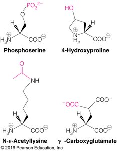
Amino acids in proteins can be modified after translation, affecting protein function, stability, and localization. Common modifications include phosphorylation, acetylation, and disulfide bond formation.
Protein Sequence Analysis and Evolution
Proteolytic Cleavage and Sequencing
Proteases and chemical reagents cleave proteins at specific sites, facilitating sequence analysis. Modern techniques include mass spectrometry and Edman degradation.
Enzyme | Preferred Site | Source |
|---|---|---|
Trypsin | R1 = Lys, Arg | Digestive systems of animals |
Chymotrypsin | R1 = Tyr, Trp, Phe, Leu | Same as trypsin |
Thrombin | R1 = Arg | Blood; coagulation |
V8 protease | R1 = Asp, Glu | Staphylococcus aureus |
Cyanogen bromide | R1 = Met | Chemical reagent |

Evolutionary Relationships
Protein sequences are compared to infer evolutionary relationships and predict function. Conservative mutations preserve chemical properties, while nonconservative mutations do not. Tools like BLAST identify sequence similarities and evolutionary conservation.
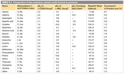
Summary Table: Properties of Common Amino Acids
Name | Abbreviations | pKa of -COOH | pKa of -NH3+ | pKa of Side Chain | Residue Mass (Da) | Occurrence (%) |
|---|---|---|---|---|---|---|
Alanine | A, Ala | 2.3 | 9.7 | - | 71.08 | 8.7 |
Arginine | R, Arg | 2.2 | 9.0 | 12.5 | 156.19 | 5.2 |
Asparagine | N, Asn | 2.1 | 8.8 | - | 114.10 | 4.2 |
Aspartic acid | D, Asp | 2.1 | 9.8 | 3.9 | 115.09 | 5.5 |
Cysteine | C, Cys | 1.7 | 10.8 | 8.3 | 103.15 | 1.9 |
Glutamine | Q, Gln | 2.2 | 9.1 | - | 128.13 | 3.9 |
Glutamic acid | E, Glu | 2.2 | 9.7 | 4.2 | 129.12 | 6.2 |
Glycine | G, Gly | 2.3 | 9.6 | - | 57.05 | 7.2 |
Histidine | H, His | 1.8 | 9.2 | 6.0 | 137.14 | 2.3 |
Isoleucine | I, Ile | 2.2 | 9.9 | - | 113.17 | 5.3 |
Leucine | L, Leu | 2.3 | 9.7 | - | 113.17 | 9.0 |
Lysine | K, Lys | 2.2 | 9.2 | 10.0 | 128.18 | 7.0 |
Methionine | M, Met | 2.1 | 9.3 | - | 131.21 | 2.3 |
Phenylalanine | F, Phe | 2.2 | 9.2 | - | 147.18 | 3.9 |
Proline | P, Pro | 2.0 | 10.6 | - | 97.12 | 4.7 |
Serine | S, Ser | 2.2 | 9.2 | - | 87.08 | 6.6 |
Threonine | T, Thr | 2.1 | 9.1 | - | 101.11 | 5.3 |
Tryptophan | W, Trp | 2.5 | 9.4 | - | 186.21 | 1.1 |
Tyrosine | Y, Tyr | 2.2 | 9.2 | 10.1 | 163.18 | 3.2 |
Valine | V, Val | 2.3 | 9.6 | - | 99.14 | 7.2 |
Additional info: This guide covers the foundational aspects of protein structure, amino acid chemistry, and protein purification, as outlined in a typical biochemistry curriculum.
