 Indietro
Protein Analysis and Proteomics: Techniques and Applications
Guida di studio - Note intelligenti
 Appunti personalizzati basati sui tuoi materiali, ampliati con definizioni chiave, esempi e contesto.
Protein Analysis: Principles and Methods
Introduction to Protein Analysis
Protein analysis is fundamental in biochemistry for understanding protein structure, function, and interactions. It encompasses a variety of techniques to determine protein concentration, separate proteins based on their properties, sequence proteins, and analyze their roles in biological systems.
Quantification of Proteins by Spectrophotometry
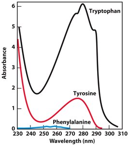
Proteins can be quantified using their absorbance in the ultraviolet (UV) region, primarily due to aromatic amino acids such as tryptophan and tyrosine. This method is rapid and non-destructive, suitable for both pure proteins and mixtures.
Key Principle: Aromatic amino acids absorb UV light maximally at 275–280 nm.
Major Chromophores: Tryptophan (strongest), tyrosine, and phenylalanine (weakest).
Alternative Assay: The Biuret assay is used for proteins lacking aromatic residues.
Protein Separation Techniques
Principles of Protein Separation
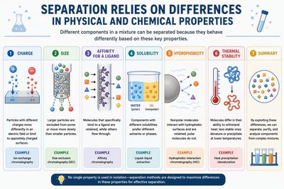
Proteins are separated based on differences in their physical and chemical properties. Effective separation is crucial for protein purification and analysis.
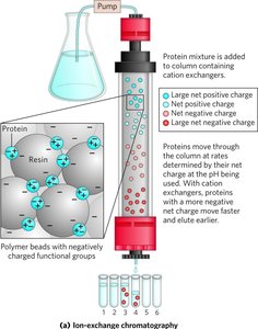
Charge: Exploited in ion-exchange chromatography.
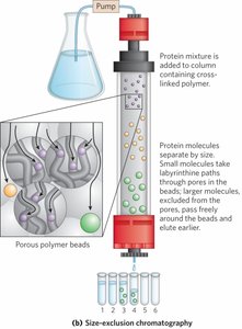
Size: Used in size-exclusion (gel filtration) chromatography.
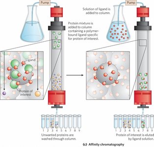
Affinity for a Ligand: Basis for affinity chromatography.
Solubility: Manipulated in selective precipitation methods.
Hydrophobicity: Used in hydrophobic interaction chromatography.
Thermal Stability: Some proteins can be separated by heat denaturation.
Selective Precipitation Methods
Effect of pH on Solubility
The solubility of proteins is highly dependent on pH, which affects their net charge and aggregation behavior.
At pH < pI: Protein has a net positive charge and is stabilized in solution.
At pH = pI: Net charge is zero, leading to aggregation and precipitation.
At pH > pI: Protein has a net negative charge and is stabilized in solution.
Protein | pI |
|---|---|
Lysozyme | 11.35 |
Ovalbumin | 4.5 |
Pepsin | <1 |
Effect of Ionic Strength on Solubility
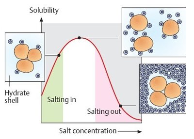
Protein solubility first increases with salt concentration ('salting in'), then decreases at high salt ('salting out'). This is due to the competition between salt ions and proteins for water molecules.
Salting in: Low salt concentrations shield protein charges, increasing solubility.
Salting out: High salt concentrations reduce available water, causing proteins to aggregate and precipitate.
Hofmeister Series: Ranks ions by their ability to precipitate proteins; ammonium sulfate is commonly used.
Effect of Organic Solvents
Adding organic solvents (e.g., acetone, ethanol) lowers the dielectric constant of water, reducing protein solubility and causing precipitation. This method is less commonly used due to the flammability of solvents.
Chromatographic Methods for Protein Separation
Column Chromatography: General Principles
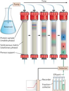
Column chromatography separates proteins based on their interactions with a stationary phase (solid matrix) and a mobile phase (liquid). Proteins are separated as they move through the column at different rates. (sepearte them beause some come out slower than others/ at different speeds)
Types of Chromatography
Size-Exclusion Chromatography (Gel Filtration)
Separates proteins based on size. Large proteins elute first as they are excluded from the pores of the matrix, while smaller proteins enter the pores and elute later.
Ion-Exchange Chromatography
Separates proteins based on net charge at a given pH. Proteins bind to oppositely charged groups on the matrix and are eluted by changing salt concentration or pH.
Affinity Chromatography
Exploits specific binding interactions between a protein and a ligand attached to the matrix. Highly selective and efficient for purifying target proteins.
Electrophoretic Techniques
Polyacrylamide Gel Electrophoresis (PAGE)
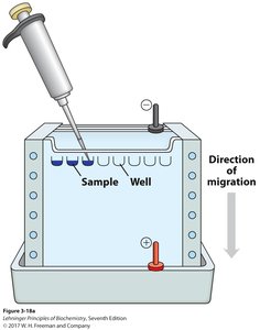
PAGE separates proteins based on their size and charge under an electric field. The gel matrix hinders movement, allowing separation by molecular weight.
Native PAGE: Maintains protein structure and separates by size and charge.
SDS-PAGE
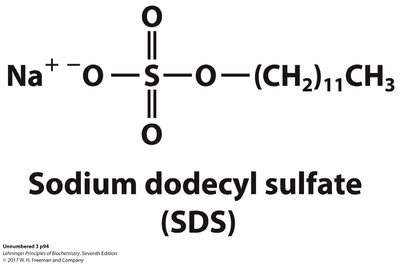
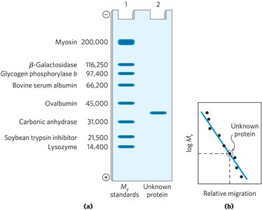
Sodium dodecyl sulfate (SDS) denatures proteins and gives them a uniform negative charge, allowing separation strictly by size. Used to determine molecular weight and assess purity.
Isoelectric Focusing (IEF)
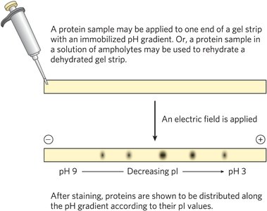
Proteins are separated in a pH gradient gel until they reach their isoelectric point (pI), where their net charge is zero and migration stops.
Two-Dimensional Gel Electrophoresis (2D-GE)
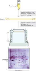
Combines IEF and SDS-PAGE for high-resolution separation. First, proteins are separated by pI, then by size, allowing analysis of complex mixtures.
 Purity and Activity
Protein
Activity vs. Specific Activity
Specific activity is the ratio of enzyme activity to total protein, indicating purity. As purification proceeds, specific activity increases.
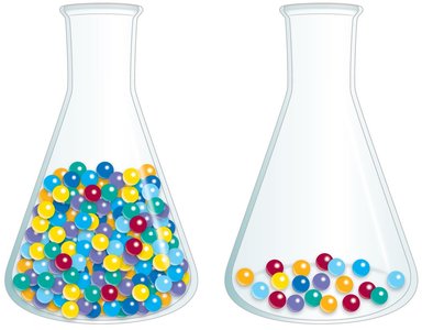
Candy analogy:
Imagine a bag with 100 candies total. Only the red candies are the “special ones” that do the job (that’s your enzyme). The other colors are just extra protein that doesn’t do the reaction.
Activity = “how much job gets done” depends on how many red candies you have. More red candies → more activity.
Total protein = “how many candies in the bag” (red + all other colors).
Specific activity = “how many red candies per total candies” (but measured as activity per mg protein). It tells you: How concentrated is the enzyme inside all the protein?
Example: Bag A: 10 red candies + 90 other candies (100 total). The bag can do some work, but it’s mostly junk → low specific activity.
Immunological Techniques
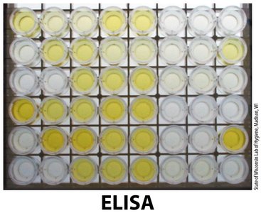
Enzyme-Linked Immunosorbent Assay (ELISA)
ELISA detects and quantifies specific proteins using antibodies. The presence of antigen-antibody complexes is revealed by a color change.
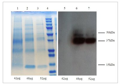
Western Blotting
Proteins separated by SDS-PAGE are transferred to a membrane, probed with specific antibodies, and visualized. Used for detection and quantification of target proteins.
Protein Sequencing and Sequence Analysis
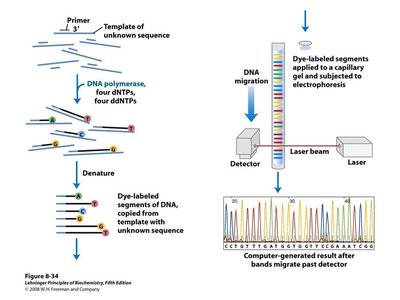
DNA Sequencing for Protein Analysis
DNA sequencing (e.g., Sanger method) allows determination of the coding sequence for proteins, revealing amino acid sequence and potential mutations.
Protein Sequence Variations and Disease
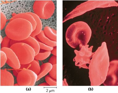
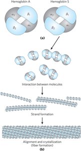
Comparing protein sequences can identify mutations linked to diseases or evolutionary relationships. For example, sickle cell anemia results from a single amino acid substitution in hemoglobin.
Proteomics
Introduction to Proteomics
Proteomics is the large-scale study of proteins, including their expression, structure, and function. It provides insights into cellular processes, disease mechanisms, and evolutionary biology.
Bottom-Up and Top-Down Proteomics
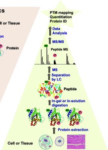
Bottom-Up: Proteins are digested into peptides before analysis by mass spectrometry.
Top-Down: Intact proteins are analyzed directly, preserving post-translational modifications.
Mass Spectrometry in Proteomics
Mass spectrometry (MS) is a powerful tool for protein identification and characterization. It provides information on protein mass, sequence, and structure.
Ionization Methods: Electrospray ionization (ESI) and matrix-assisted laser desorption ionization (MALDI).
Mass Analyzers: Time-of-flight (TOF), quadrupole, and triple quadrupole (QQQ).
Applications of Proteomics
Systems Biology: Understanding cellular pathways and networks.
Biomarker Discovery: Identifying disease markers for diagnostics and therapy monitoring.
Drug Target Evaluation: Assessing protein targets for drug development and toxicity studies.
