Indietro
Protein Function: Ligand Binding, Immune Proteins, and Molecular Motors
Guida di studio - Note intelligenti
 Appunti personalizzati basati sui tuoi materiali, ampliati con definizioni chiave, esempi e contesto.
Protein Function
Introduction to Protein-Ligand Interactions
Proteins perform a wide range of functions in biological systems, many of which depend on their ability to bind other molecules, called ligands, reversibly and with high specificity. The nature of these interactions underlies processes such as oxygen transport, immune defense, and muscle contraction.
Ligand: A molecule that binds specifically to a protein at its binding site (often called a pocket).
Affinity: The strength of the binding interaction between a protein and its ligand.
Specificity: The ability of a protein to selectively bind a particular ligand among many possible molecules.
Overview of Protein–Ligand Interactions
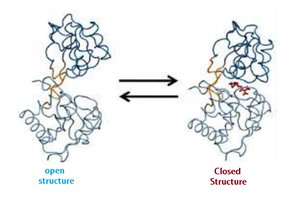
Protein-ligand interactions are governed by several factors, including shape complementarity, charge interactions, and hydrophobic/hydrophilic effects. The classic lock-and-key model describes how a ligand fits into a protein's binding site, but many proteins also undergo conformational changes upon ligand binding (induced fit).
Induced Fit: The protein changes shape to better accommodate the ligand, enhancing binding affinity and specificity.
Example: G-protein-coupled receptors (GPCRs) and maltose-binding protein (MBP) undergo significant conformational changes upon ligand binding.
Allosteric Proteins and Modulation
Some proteins have multiple binding sites and can be regulated by ligands binding at sites other than the active site (allosteric regulation). Modulators can be activators or inhibitors, and their effects can be homotropic (same molecule as the ligand) or heterotropic (different molecule).
Allosteric Protein: Binding of a ligand at one site affects binding properties at another site.
Homotropic Interactions: Multiple molecules of the same ligand bind cooperatively.
Heterotropic Interactions: Modulator is a different molecule than the normal ligand.
Oxygen-Binding Proteins: Hemoglobin and Myoglobin
Globin Family and Hemoglobin Structure
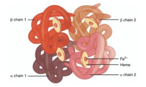
The globin family includes proteins such as hemoglobin and myoglobin, which are essential for oxygen transport and storage. Hemoglobin is a tetrameric protein found in red blood cells, while myoglobin is a monomeric protein found in muscle tissue.
Hemoglobin: Composed of four subunits, each containing a heme group that binds oxygen.
Heme Group: A prosthetic group containing Fe2+ at its center, essential for reversible oxygen binding.
Heme Structure and Protein Association
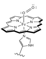
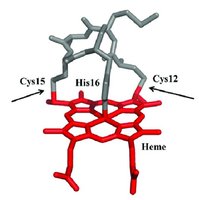
The heme group is a planar structure with an iron ion coordinated by nitrogen atoms of a porphyrin ring and a histidine side chain from the protein. This arrangement protects the iron from oxidation and allows reversible oxygen binding.
Protoporphyrin: The organic component of heme.
Histidine Coordination: The iron is held in place by a histidine residue, which is crucial for function.
Protein-Ligand Heme Association Motif
Heme is covalently attached to proteins via a CXXCH motif in some proteins, such as c-type cytochromes, which are involved in electron transfer.
CXXCH Motif: Sequence motif where heme is covalently linked to cysteine residues and coordinated by histidine.
Reversible Binding and Quantitative Analysis
Most protein-ligand interactions are non-covalent, involving hydrogen bonds, ionic interactions, van der Waals forces, and hydrophobic effects. This allows for dynamic binding and release, essential for biological function.
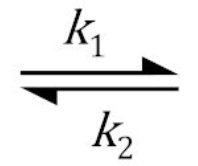
Binding Reaction:
Equilibrium: Characterized by association () and dissociation () rate constants.
Quantitative Measures: Dissociation Constant and Hill Equation
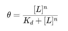
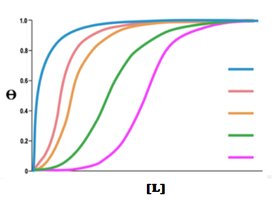
The strength of protein-ligand binding is described by the dissociation constant (). Cooperative binding, as seen in hemoglobin, is described by the Hill equation.
Dissociation Constant (): Lower indicates higher affinity.
Hill Equation: where is the fraction of occupied binding sites, is the Hill coefficient (degree of cooperativity).
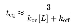
Time to Equilibrium
The time required to reach binding equilibrium depends on ligand concentration and rate constants. For high ligand concentrations, equilibrium is reached rapidly.
Approximate Time to Equilibrium:
Regulated Ligand Binding
In multi-subunit proteins, ligand binding to one subunit can affect the binding of additional ligands to other subunits, a phenomenon known as cooperativity. Aspartate transcarbamoylase (ATCase) is a classic example, regulated by both positive and negative effectors.
Positive Cooperativity: Binding of one ligand increases affinity for subsequent ligands.
Negative Regulation: End products (e.g., CTP) inhibit enzyme activity via feedback inhibition.
Immune System Proteins: Antibodies and Immunoglobulins
Immune Response and Immunoglobulins
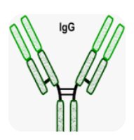
The immune system relies on a variety of proteins, including antibodies (immunoglobulins), to recognize and neutralize foreign molecules (antigens). Immunoglobulins are classified into several types, with IgG being the most abundant in blood serum.
Antigen: A molecule recognized by the immune system.
Epitope: The specific part of an antigen recognized by an antibody.
Immunoglobulins: Proteins that bind antigens with high specificity.
Antibody Structure and Domains
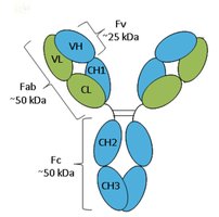
Antibodies are Y-shaped molecules composed of two heavy and two light chains, each containing variable and constant domains. The variable domains confer antigen specificity, while the constant domains mediate immune effector functions.
Immunoglobulin Fold: A β-sandwich structure formed by two β-sheets.
Fab Region: Contains antigen-binding sites (VH, VL, CH1, CL).
Fc Region: Responsible for effector functions (CH2, CH3).
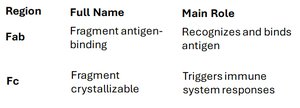
Region | Full Name | Main Role |
|---|---|---|
Fab | Fragment antigen-binding | Recognizes and binds antigen |
Fc | Fragment crystallizable | Triggers immune system responses |
IgM Structure and Function
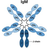
IgM is a pentameric antibody, providing strong initial immune responses due to its high avidity. It contains a J chain required for polymerization and transport across mucosal surfaces.
Pentameric Structure: Five IgM monomers joined by a J chain, with 10 antigen-binding sites.
Fc Region: Central cluster of CH2, CH3, and CH4 domains.
Protein Interactions Modulated by Chemical Energy: Actin, Myosin, and Molecular Motors
Muscle Proteins: Actin and Myosin
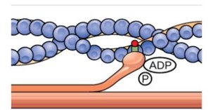
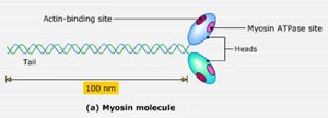
Muscle contraction is driven by the interaction of actin and myosin filaments, powered by ATP hydrolysis. Myosin heads bind to actin, undergo conformational changes, and generate force for contraction.
Actin Filament: Provides the track for myosin movement.
Myosin: Motor protein with ATPase activity, responsible for force generation.
Regulation of Muscle Contraction
Muscle contraction is regulated by the binding of Ca2+ ions to troponin, which causes a conformational change in tropomyosin, exposing the actin-myosin binding site and allowing contraction to proceed.
Troponin: Binds Ca2+ and regulates tropomyosin position.
Tropomyosin: Blocks actin-myosin interaction in the absence of Ca2+.