 Indietro
Protein Function: Reversible Ligand Binding and Immunoglobulins
Guida di studio - Note intelligenti
 Appunti personalizzati basati sui tuoi materiali, ampliati con definizioni chiave, esempi e contesto.
Protein Function: Reversible Ligand Binding
Introduction to Protein-Ligand Binding
Proteins perform their biological functions by interacting dynamically with other molecules, often referred to as ligands. These interactions are fundamental to processes such as signaling, transport, and immune response. - Ligand: A molecule that binds specifically to a protein, often resulting in a functional change. - Reversible binding: Most protein-ligand interactions are reversible, allowing for regulation and control of biological processes. 
Types of Protein Interactions
Proteins interact with other molecules in two primary ways: - Transient interactions: Short-lived, such as enzyme-substrate binding. - Stable interactions: Longer-lasting, such as structural protein complexes.
Oxygen-Binding Proteins: Globins
Globins as Oxygen-Binding Proteins
Globins are a family of proteins specialized for binding and transporting oxygen. - Myoglobin: Found in muscle tissue, stores oxygen for use during intense activity. - Hemoglobin: Found in red blood cells, transports oxygen from lungs to tissues. 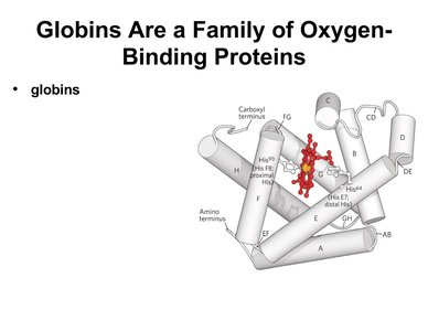
Myoglobin: Single Oxygen Binding Site
Myoglobin contains a single binding site for oxygen, making it efficient for oxygen storage but not for transport. - Structure: Myoglobin is a monomeric protein with a heme prosthetic group. 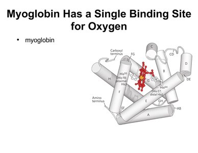
Oxygen Binding to Heme Prosthetic Group
Oxygen binds to the iron atom in the heme group, which is embedded within the globin protein. - Heme: A prosthetic group consisting of an iron ion coordinated within a porphyrin ring. - Function: Enables reversible oxygen binding. 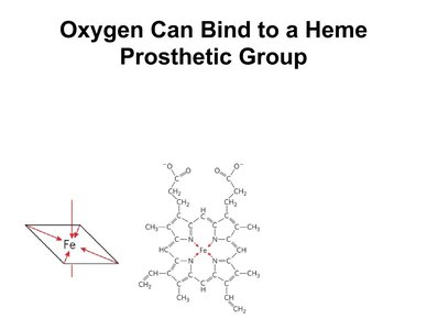
Quantitative Description of Protein-Ligand Interactions
Equilibrium Expression for Binding
The reversible binding of a protein (P) to a ligand (L) can be described by an equilibrium expression: - Equation: - Equilibrium constant (Ka): - Dissociation constant (Kd):
Graphical Representations of Ligand Binding
Ligand binding is often visualized using binding curves, which plot the fraction of occupied binding sites versus ligand concentration.
Hemoglobin Structure and Function
Structural Similarity of Hemoglobin and Myoglobin
Hemoglobin subunits are structurally similar to myoglobin, each containing a heme group. - Hemoglobin: Tetrameric protein with two alpha and two beta subunits. 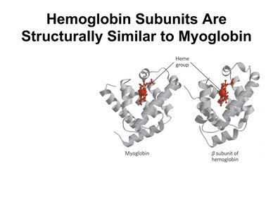
Hemoglobin Transports Oxygen in Blood
Hemoglobin is responsible for oxygen transport in the circulatory system. - Transport mechanism: Binds oxygen in the lungs, releases it in tissues.
Quaternary Structure of Hemoglobin
Hemoglobin exhibits quaternary structure, consisting of four polypeptide chains. - Subunits: Two alpha and two beta chains. - Heme groups: Each subunit contains a heme group. 
Cooperative Oxygen Binding
Hemoglobin binds oxygen cooperatively, meaning the binding of one oxygen molecule increases the affinity for subsequent oxygen molecules. - Cooperativity: Allows efficient oxygen loading and unloading. 
Structural Changes Upon Oxygen Binding
Hemoglobin undergoes a conformational change from the T (tense) state to the R (relaxed) state upon oxygen binding. - T state: Lower affinity for oxygen. - R state: Higher affinity for oxygen. 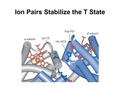
Ion Pairs Stabilize the T State
Ion pairs between subunits stabilize the T state, contributing to the regulation of oxygen affinity. 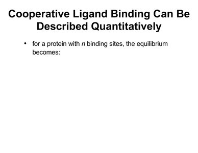
Quantitative Description of Cooperative Binding
For proteins with multiple binding sites, the equilibrium expression is modified: - Hill equation: where is the fraction of occupied sites, is ligand concentration, is the Hill coefficient.
Hill Plots and Hill Coefficients
Hill plots are used to analyze cooperativity in ligand binding. - Hill coefficient (n): Indicates the degree of cooperativity; suggests positive cooperativity.
Sickle Cell Anemia: Molecular Disease of Hemoglobin
Pathogenesis of Sickle Cell Anemia
Sickle cell anemia is caused by a mutation in hemoglobin, leading to abnormal aggregation and deformation of red blood cells. - Mutation: Substitution of valine for glutamic acid in the beta chain. - Effect: Hemoglobin S polymerizes under low oxygen, distorting cell shape. 
Complementary Interactions: Immune System and Immunoglobulins
Antibodies and Antigen Binding
Antibodies (immunoglobulins) are proteins that bind specifically to antigens, facilitating immune response. - Structure: Y-shaped molecules with two identical antigen-binding sites. 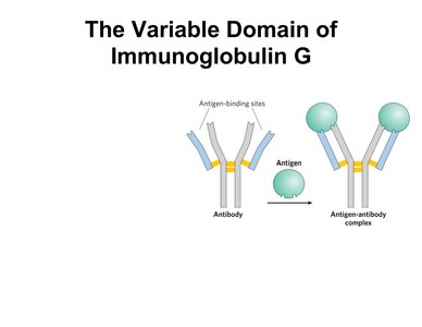
Variable Domain of Immunoglobulin G
The variable domain of IgG determines antigen specificity. - Antigen-binding sites: Located at the tips of the Y-shaped molecule. 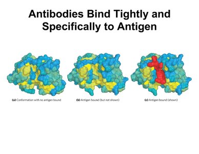
Antibody-Antigen Binding Specificity
Antibodies bind tightly and specifically to their target antigens, enabling precise immune recognition. Example: Immunoglobulin G binding to a viral protein, neutralizing its activity.
Additional info: Academic context was added to clarify quantitative binding expressions, structural details, and immune system relevance.
